Home > Science > SEM
Eyebrow hair, SEM
![]()

Wall Art and Photo Gifts from Science Photo Library
Eyebrow hair, SEM
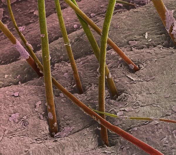
Eyebrow hair and skin. Coloured scanning electron micrograph (SEM) of eyebrow hair growing from the surface of human skin. The shafts of hair (brown) are anchored in their individual hair follicles (not seen) below the surface of the skin (pink). Hair is made up of a fibrous protein called keratin. The outermost skin layer, the stratum corneum, consists of keratinized dead cells that detach from the body. The squamous (flattened) cells that make up the stratum corneum arise from the lower, living layers of skin. Magnification: x100 at 6x7cm size
Science Photo Library features Science and Medical images including photos and illustrations
Media ID 6455727
© STEVE GSCHMEISSNER/SCIENCE PHOTO LIBRARY
Dead Epidermal Epidermis Eye Brow Facial Follicle Follicles Hair Hairs Histology Keratin Keratinized Magnified Image Microscopic Photos Shaft Shafts Skin Squamous Stratum Corneum Subjects Surface Tissue Cells
FEATURES IN THESE COLLECTIONS
EDITORS COMMENTS
This print from Science Photo Library offers a mesmerizing glimpse into the intricate world of eyebrow hair and skin. Captured using a scanning electron microscope (SEM), the image showcases the delicate strands of eyebrow hair emerging from the surface of human skin. The brown shafts of hair, anchored in their individual follicles beneath the pink-hued skin, are beautifully highlighted in this colored SEM. It is fascinating to note that these hairs are composed primarily of keratin, a fibrous protein found abundantly throughout our bodies. The outermost layer of skin, known as the stratum corneum, consists mainly of keratinized dead cells that naturally detach from our bodies over time. The squamous or flattened cells forming this layer originate from deeper layers within our living epidermis. At a magnification level of x100 on a 6x7cm print size, this microscopic photo provides an up-close look at healthy tissue and normal anatomy. Its scientific significance lies not only in its exploration of facial features but also in shedding light on histology and epidermal structures. Science enthusiasts and researchers alike will appreciate this visually stunning depiction that delves into subjects such as cell biology and human body composition. This particular photograph should not be used for commercial purposes without proper authorization; however, it serves as an invaluable educational resource for those interested in understanding the complexity and beauty hidden within even seemingly mundane aspects like eyebrow hair growth.
MADE IN THE UK
Safe Shipping with 30 Day Money Back Guarantee
FREE PERSONALISATION*
We are proud to offer a range of customisation features including Personalised Captions, Color Filters and Picture Zoom Tools
SECURE PAYMENTS
We happily accept a wide range of payment options so you can pay for the things you need in the way that is most convenient for you
* Options may vary by product and licensing agreement. Zoomed Pictures can be adjusted in the Basket.

