Home > Science > SEM
Coloured SEM of section through human spleen

![]()
Wall Art and Photo Gifts from Science Photo Library
Coloured SEM of section through human spleen
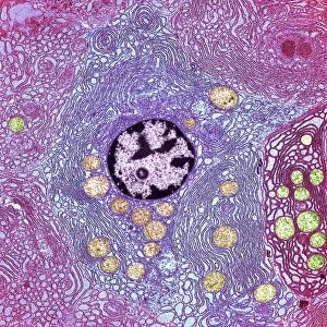
Human spleen. Coloured Scanning Electron Micro- graph (SEM) of a section through the human spleen. Here, the fine network of reticular fibres known as trabeculae is seen. Trabeculae form a framework within which the functions of the spleen take place. The spleen is part of the lymphatic system, and is involved in processing white blood cells (lymphocytes). Lymphocytes occur in round nodules, of which the framework of a nodule is seen at centre. Between the nodules there is a matrix containing red pulp, made up largely of red blood cells. The spleen also regulates the number of red blood cells in circulation in the bloodstream. Magnification: x4000 at 6x4.5cm size
Science Photo Library features Science and Medical images including photos and illustrations
Media ID 6446957
© PASIEKA/SCIENCE PHOTO LIBRARY
Magnified Image Microscopic Photos Spleen Subjects Trabeculae
FEATURES IN THESE COLLECTIONS
EDITORS COMMENTS
This print showcases a coloured scanning electron micrograph (SEM) of a section through the human spleen. The intricate details revealed in this image are truly awe-inspiring. At the heart of the composition, we can observe the fine network of reticular fibres known as trabeculae, which form a vital framework within which the functions of the spleen take place. As an integral part of the lymphatic system, the spleen plays a crucial role in processing white blood cells called lymphocytes. These lymphocytes are organized into round nodules, and at the center of this image, we catch a glimpse of their fascinating framework. In between these nodules lies a matrix containing red pulp predominantly composed of red blood cells. Not only does this remarkable organ process and regulate white blood cells but it also has control over red blood cell count in circulation throughout our bloodstream. This microscopic view offers us an extraordinary insight into how our bodies function on such minute levels. With its magnification set at x4000 and presented in 6x4.5cm size, this photograph from Science Photo Library is not just visually stunning but also serves as an educational tool for anyone interested in anatomy or exploring subjects related to human body structures under high magnification microscopy.
MADE IN THE UK
Safe Shipping with 30 Day Money Back Guarantee
FREE PERSONALISATION*
We are proud to offer a range of customisation features including Personalised Captions, Color Filters and Picture Zoom Tools
SECURE PAYMENTS
We happily accept a wide range of payment options so you can pay for the things you need in the way that is most convenient for you
* Options may vary by product and licensing agreement. Zoomed Pictures can be adjusted in the Basket.