Fraud Blocker Skip to main content

Fungal Collection (page 9)

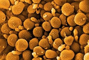
"Fungal Kingdom: Exploring the Intricate World of Fungi" Budding yeast cell: Witness the remarkable process of reproduction as a budding yeast cell emerges

Background imageFungal Collection: Bracket fungus (Daedaleopsis confragosa)

Bracket fungus (Daedaleopsis confragosa)
Blushing bracket fungus (Daedaleopsis confragosa). Close-up of the underside of the blushing bracket fungus showing its pores. The pores are the ends of the tubes within which the spores are produced

Background imageFungal Collection: Bracket fungus (Polyporus squamosus)

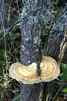
Bracket fungus (Polyporus squamosus)
Bracket fungus. Dryads saddle (Polyporus squamosus) bracket fungus on a tree

Background imageFungal Collection: Parasol mushrooms

Parasol mushrooms (Macrolepiota procera). Photographed in Narke, Sweden

Background imageFungal Collection: Psathyrella mushrooms after forest fire

Psathyrella mushrooms after forest fire
Psathyrella pennata mushrooms growing one year after a forest fire. This fire took place in a clear-felled area in the Tiveden forest, in Vastra Gotaland County, Sweden

Background imageFungal Collection: Geopyxis elf-cup fungi after forest fire

Geopyxis elf-cup fungi after forest fire
Geopyxis carbonaria elf-cup fungi growing one year after a forest fire. This fire took place in a clear-felled area in the Tiveden forest, in Vastra Gotaland County, Sweden

Background imageFungal Collection: Reishi fungus

Reishi fungus
Reishe fungus (Ganoderma lucidum) growing on a dead tree. Photographed in Narke, Sweden

Background imageFungal Collection: Honey fungus

Honey fungus (Armillaria mellea) growing on a tree trunk. Photogroahed in Narke, Sweden

Background imageFungal Collection: Jersey cow bolete

Jersey cow bolete (Suillus bovinus). Photographed in Narke, Sweden

Background imageFungal Collection: Myxomphalia mushroom after forest fire

Myxomphalia mushroom after forest fire
Myxomphalia maura mushrooms growing one year after a forest fire. This fire took place in a clear-felled area in the Tiveden forest, in Vastra Gotaland County, Sweden

Background imageFungal Collection: Helvella macropus mushroom

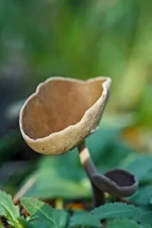
Helvella macropus mushroom
Helvella macropus fungus fruiting bodies. Photographed in Narke, Sweden

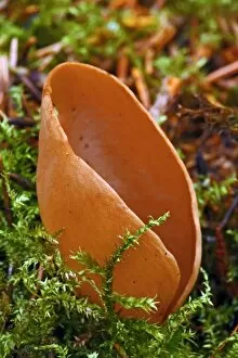
Background imageFungal Collection: Hares ear fungus

Hares ear fungus (Otidea onotica). Photographed in Narke, Sweden

Background imageFungal Collection: Maze-gill fungus underside

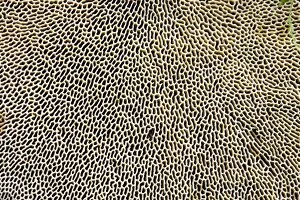
Maze-gill fungus underside
Maze-gill fungus (Daedalea quercina) underside, showing the gills where the reproductive spores (not seen) are released. Photographed in October, on Powerstock Common, Dorset, UK

Background imageFungal Collection: Peziza cup fungus after forest fire

Peziza cup fungus after forest fire
Peziza cup fungus (Peziza lobulata) growing two months after a forest fire. This fire took place in a clear-felled area in the Tiveden forest, in Vastra Gotaland County, Sweden

Background imageFungal Collection: Plant regrowth after forest fire

Plant regrowth after forest fire
Plant regrowth one year after a forest fire. The plants and fungi seen here include Pholiota highlandensis mushrooms (lower left), the common liverwort (Marchantia polymorpha, white-green, centre)

Background imageFungal Collection: Tar Spot Fungus (Rhytisma acerinum)

Tar Spot Fungus (Rhytisma acerinum) on the leaves of a Field Maple (Acer campestre) tree. Photographed in autumn in Romania

Background imageFungal Collection: Birch bolete (Leccinum scabrum) mushroom

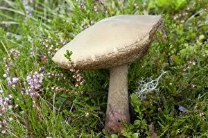
Birch bolete (Leccinum scabrum) mushroom
Brown birch bolete (Leccinum scabrum) mushroom growing amongst common heather (Calluna vulgaris). Photographed in the Cairngorms National Park, Scotland, in August

Background imageFungal Collection: Lichen (Sticta sylvatica)

Lichen (Sticta sylvatica) growing on rocks. Photographed in Auvergne in France

Background imageFungal Collection: Bean rust pustules, SEM

Bean rust pustules, SEM
Bean rust (Uromyces sp.) pustules, coloured scanning electron micrograph (SEM). The spores (orange) are emerging from pustules on the underside of a leaf on a bean plant

Background imageFungal Collection: Mushroom gill cap, light micrograph

Mushroom gill cap, light micrograph. Transverse section through the cap of a common mushroom (Psalliota sp.) showing the gill lamellae (white)

Background imageFungal Collection: Bean rust spores, SEM

Bean rust spores, SEM
Bean rust (Uromyces sp.) spores, coloured scanning electron micrograph (SEM). Bean rust is a fungal disease that infects bean plants

Background imageFungal Collection: Rust fungus infection, light micrograph

Rust fungus infection, light micrograph. Transverse section through a barberry (Berberis sp.) leaf infected by a parasitic rust (Puccinia sp.)

Background imageFungal Collection: Fly agaric (Amanita muscaria) mushrooms

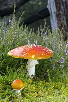
Fly agaric (Amanita muscaria) mushrooms growing amongst common heather (Calluna vulgaris) in an ancient Caledonian Scots Pine woodland

Background imageFungal Collection: Mould fruiting bodies, light micrograph

Mould fruiting bodies, light micrograph. Black mould (Rhizopus nigricans) fruiting bodies (top and upper right) liberating spores

Background imageFungal Collection: Aspergillus fungus spores, SEM

Aspergillus fungus spores, SEM
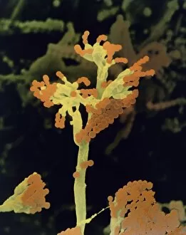
Aspergillus fungus spores, coloured scanning electron micrograph (SEM). These chains of spores (orange) are the reproductive cells of the fruiting body, or conidiophore

Background imageFungal Collection: Penicillium fungus, SEM

Penicillium fungus, SEM
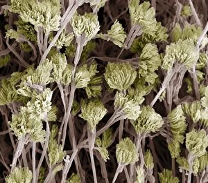
Penicillium fungus. Coloured scanning electron micrograph (SEM) of a Penicillium sp. fungus. Specialised threads (hyphae, pink), called conidiophores, are seen

Background imageFungal Collection: Amanita (Amanita frostiana) mushroom

Amanita (Amanita frostiana) mushroom
Frosts amanita (Amanita frostiana) mushroom. Photographed in Vermont, USA

Background imageFungal Collection: Woolly gomphus (Gomphus floccosus)

Woolly gomphus (Gomphus floccosus) mushroom. Photographed in Vermont, USA

Background imageFungal Collection: Root fungus, light micrograph

Root fungus, light micrograph
Root fungus. Light micrograph of a cross-section through a root containing fungal hyphae called endotrophic mycorrhiza. The hyphae (green and blue)

Background imageFungal Collection: Japanese sago palm root, light micrograph

Japanese sago palm root, light micrograph
Japanese sago palm root. Light micrograph of a transverse section through a root of a Japanese sago palm, (Cycas revoluta)

Background imageFungal Collection: Edible fungi at a market

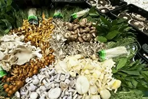
Edible fungi at a market. Selection of different mushrooms and fungal fruiting bodies arrayed for sale on a market stall. There are thousands of species of edible mushrooms found worldwide

Background imageFungal Collection: Dry rot fungus

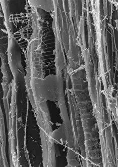
Dry rot fungus. Dry rot, unlike its name suggests, infects damp timber. The term refers to the growth of either of two fungi

Background imageFungal Collection: Macrophoto of penicillium roqueforti

Macrophoto of penicillium roqueforti
Macrophotograph (filtered) of Penicillium roqueforti, an economically important penicillin fungus used in the manufacture of blue cheeses

Background imageFungal Collection: Lace lichen (Ramalina menziesii)

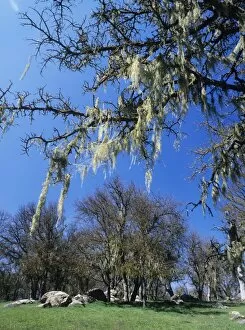
Lace lichen (Ramalina menziesii) growing on the branches of a valley oak tree (Quercus lobata). Lichens are symbiotic organisms formed of a fungus and an alga that mutually benefit one another

Background imageFungal Collection: Photomicrograph of Penicillin chrysogenum

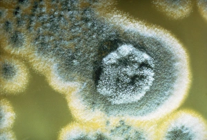
Photomicrograph of Penicillin chrysogenum, 3 days old, grown on potato dextrose agar medium (yellow). Magnification: X 1.2 at 35mm size

Background imageFungal Collection: Sulphur toadstools

Sulphur toadstools. Undersides of sulphur toadstools (Tricholoma sulphureum). The gills contain million of spores, the reproductive cells of a fungus

Background imageFungal Collection: SEM of dry rot in plywood

SEM of dry rot in plywood

Background imageFungal Collection: Bleeding mycena mushrooms

Bleeding mycena mushrooms (Mycena haemotopus). Bleeding mycenas are so called because they release a reddish juice when cut. They grow on rotting wood, reaching about 3-8 centimetres in height

Background imageFungal Collection: Cryptococcus cell dividing, TEM

Cryptococcus cell dividing, TEM
Cryptococcus cell dividing. Coloured transmission electron micrograph (TEM) of the asexual budding (lower right) of a Cyptococcus sp. yeast cell

Background imageFungal Collection: Daedaleopsis confragosa fungus

Daedaleopsis confragosa fungus attached to the trunk of a tree. This bracket fungus grows on dead and living wood of broad leaved trees, particularly alder, birch and willow

Background imageFungal Collection: Earth star mushroom

Earth star mushroom (Geastrum triplex) growing in woodland soil. It has released (puffed) a cloud of reproductive spores (upper centre)

Background imageFungal Collection: Lichens

Lichens

Background imageFungal Collection: False col SEM of rhizoids

False col SEM of rhizoids
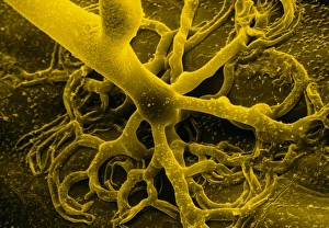
False colour scanning electron micrograph of rhizoids, or " rootlets" of the bread mould, Rhizopus stolonifer, on the surface of bread. Spores circulate freely in the air

Background imageFungal Collection: False colour SEM of fruiting bodies

False colour SEM of fruiting bodies

Background imageFungal Collection: Coloured TEM of a yeast cell

Coloured TEM of a yeast cell

Background imageFungal Collection: Puffball fungus

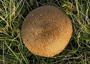
Puffball fungus (Lycoperdon foetidum)

Background imageFungal Collection: Fly agaric fungus

Fly agaric fungus (Amanita muscaria). The mycelium of the fly agaric fungus shares a symbiotic relationship with the spruce, forming a mycorrhizal sheath around the roots of the tree

Background imageFungal Collection: Shaggy parasol mushrooms

Shaggy parasol mushrooms
Shaggy parasol mushroom (Macrolepiota rhacodes). This fungus is edible

Background imageFungal Collection: Mushroom surface, SEM

Mushroom surface, SEM
Mushroom surface. Coloured scanning electron micrograph (SEM) of the spore-bearing surface of a hen of the woods mushroom (Grifola frondosa). This surface is found on the underside of the mushroom


All Professionally Made to Order for Quick Shipping

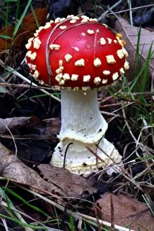
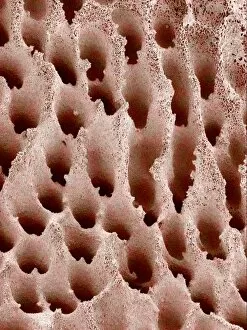
"Fungal Kingdom: Exploring the Intricate World of Fungi" Budding yeast cell: Witness the remarkable process of reproduction as a budding yeast cell emerges, ready to create new life. Fly agaric mushrooms: Delve into the enchanting realm of fly agaric mushrooms, with their vibrant red caps and white spots, captivating both nature enthusiasts and fairytale lovers alike. SEM of penicillin fungus: Uncover the microscopic beauty of penicillin fungus through scanning electron microscopy (SEM), revealing its intricate structures that have revolutionized medicine. Culture of Aspergillus nidulans fungus: Step into a laboratory where cultures of Aspergillus nidulans fungus thrive, showcasing their unique growth patterns and potential for scientific research. Cep mushroom (Boletus edulis): Discover the culinary delight known as cep mushroom or Boletus edulis, renowned for its rich flavor and sought-after by chefs around the world. Candida fungus, SEM: Explore the unseen world within our bodies as Candida fungi are magnified under an electron microscope, shedding light on their role in various infections and diseases. Mushroom gills, SEM: Marvel at the intricate network of gills found beneath mushroom caps when observed through scanning electron microscopy (SEM), highlighting their crucial role in spore production. Fly agaric fungi: Journey through enchanted forests where fly agaric fungi flourish; these iconic red-and-white toadstools evoke a sense of wonderment while harboring fascinating ecological relationships with other organisms. Dividing yeast cells, SEM: Peer into a microscopic universe where dividing yeast cells undergo mitosis under high-resolution imaging techniques—witnessing life's continuous cycle unfold before your eyes. Penicillium roqueforti fungus: Meet Penicillium roqueforti—a blue mold used in cheese-making—whose distinctive flavor and appearance contribute to the creation of beloved blue cheeses worldwide.