Fraud Blocker Skip to main content

Fungal Collection (page 13)

"Fungal Kingdom: Exploring the Intricate World of Fungi" Budding yeast cell: Witness the remarkable process of reproduction as a budding yeast cell emerges

Background imageFungal Collection: Purple coral fungus

Purple coral fungus (Clavaria zollingeri). This rare club fungus is found in unfertilised grassland habitats. It is a priority species in the Northern Ireland Biodiversity Action Plan

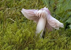
Background imageFungal Collection: Pink waxcap (Hygrocybe calyptriformis)

Pink waxcap (Hygrocybe calyptriformis)
Pink waxcap fungus (Hygrocybe calpyptriformis) amongst moss. This fungus has become rare in the UK. This is thought to be due to improvements to grassland habitats through the addition of fertiliser

Background imageFungal Collection: Magpie ink cap fungus

Magpie ink cap fungus (Coprinus picaceus). This fungus prefers alkaline soils. Photographed in woodland on the North Downs, Kent, UK, in October

Background imageFungal Collection: Honey fungus (Armillaria mellea)

Honey fungus (Armillaria mellea)
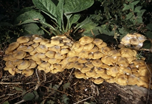
Honey fungus mushrooms (Armillaria mellea). This fungus is found on or around the trunks of coniferous and deciduous trees. It is a tree parasite and the damage it causes is fatal

Background imageFungal Collection: Orange birch bolete fungus

Orange birch bolete fungus (Leccinum versipelle). This structure is the fruiting body (mushroom) of the fungus. It is surrounded by marsh hair moss (Polytrichum commune)

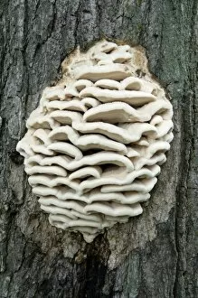
Background imageFungal Collection: Bracket fungus

Bracket fungus (class Basidiomycetes). This fungus forms these fruiting bodies to spread its reproductive spores through surface pores. Photographed in Vermont, USA

Background imageFungal Collection: Porcelain mushrooms

Porcelain mushrooms (Oudemansiella mucida) growing on a moss-covered branch in October. Photographed in Cornwall, UK

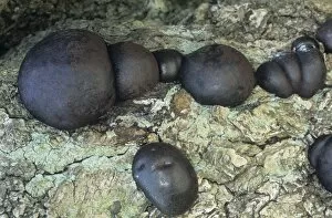
Background imageFungal Collection: King Alfreds cakes fungi

King Alfreds cakes fungi (Daldinia concentrica) growing on an ash tree (Fraxinus sp.)

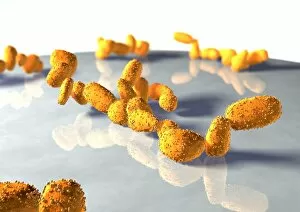
Background imageFungal Collection: Yeast cells

Yeast cells, computer artwork. Yeasts are single- cell fungi. Some yeasts have industrial uses, for example in brewing and baking, others are human pathogens

Background imageFungal Collection: Bread mould

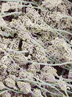
Bread mould (Penicillium sp.). Coloured scanning electron micrograph (SEM) of mouldy bread. The fungus penetrates the bread, digesting and absorbing its nutrients

Background imageFungal Collection: Honey fungus mushrooms

Honey fungus mushrooms or bootlace mushrooms (Armillaria mellea). This fungus is found on or around the trunks of coniferous and deciduous trees

Background imageFungal Collection: Fungus (Lepista caespitose)

Fungus (Lepista caespitose). Photographed in Russia

Background imageFungal Collection: Fungal spores, SEM

Fungal spores, SEM
Fungal spores. Coloured scanning electron micrograph (SEM) of fungal spores on a fruiting body of an Aspergillus sp. fungus. The name for such a fruiting body is a conidiophore

Background imageFungal Collection: Bread mould, SEM

Bread mould, SEM
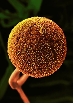
Bread mould. Coloured scanning electron micrograph (SEM) of a sporangium of a bread mould (Rhizopus stolonifer). Spores develop in these sac-like bodies, or sporangia (globular structure)

Background imageFungal Collection: Mushroom stem surface, SEM

Mushroom stem surface, SEM
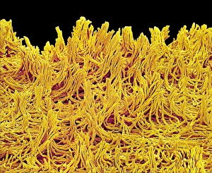
Mushroom stem. Coloured scanning electron micrograph (SEM) of the surface of the stem of an unidentified mushroom. Magnification unknown

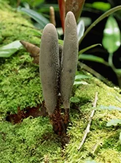
Background imageFungal Collection: Fruiting bodies of fungus, Cordyceps

Fruiting bodies of fungus, Cordyceps
Parasitic fungus. Fruiting bodies from a parasitic fungus of the Cordyceps sp. Three brown club-like fruiting bodies are seen emerging from the forest ground

Background imageFungal Collection: Parasol mushrooms, Lepiota procera

Parasol mushrooms, Lepiota procera
Parasol mushrooms. The fruiting bodies of Lepiota procera, the Parasol mushroom. This large mushroom produces a flattened pale brown cap of up to 25cm diameter

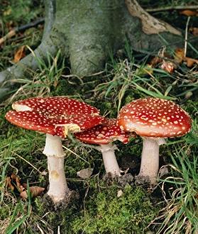
Background imageFungal Collection: Fly agaric mushrooms in wood

Fly agaric mushrooms in wood
The common mushroom known as Fly agaric, Amanita muscaria, photographed in woodland. The cap (8-20cm across) is covered with white pyramidal warts which may be washed off by rain

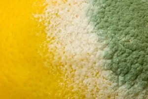
Background imageFungal Collection: Mould growing on a lemon

Mould growing on a lemon
Ashley Cooper / SpecialistStock

Background imageFungal Collection: Bread mould (Penicillium sp. ), SEM

Bread mould (Penicillium sp. ), SEM
Bread mould. Coloured scanning electronmicrograph (SEM) of Penicillium sp. mouldgrowing on bread. The yellow fibrous structuresare hyphae, which make up the main body of thefungus

Background imageFungal Collection: Mushroom

Mushroom
Close up of a fungi Mushroom

Background imageFungal Collection: Fly Agaric

Fly Agaric mushroom (Amanita muscaria) inleaf litter. This poisonous fungus, which has acap up to 20 centimetres across, is so namedbecause in medieval times it was mixed with milkand used to stupefy

Background imageFungal Collection: Bay Boletus

Bay Boletus (Boletus badius) on a woodlandfloor. Found in mixed woodland throughout theBritish Isles, the Bay Boletus is an edibleautumnal fungus which often clusters around ScotsPine trees


All Professionally Made to Order for Quick Shipping

"Fungal Kingdom: Exploring the Intricate World of Fungi" Budding yeast cell: Witness the remarkable process of reproduction as a budding yeast cell emerges, ready to create new life. Fly agaric mushrooms: Delve into the enchanting realm of fly agaric mushrooms, with their vibrant red caps and white spots, captivating both nature enthusiasts and fairytale lovers alike. SEM of penicillin fungus: Uncover the microscopic beauty of penicillin fungus through scanning electron microscopy (SEM), revealing its intricate structures that have revolutionized medicine. Culture of Aspergillus nidulans fungus: Step into a laboratory where cultures of Aspergillus nidulans fungus thrive, showcasing their unique growth patterns and potential for scientific research. Cep mushroom (Boletus edulis): Discover the culinary delight known as cep mushroom or Boletus edulis, renowned for its rich flavor and sought-after by chefs around the world. Candida fungus, SEM: Explore the unseen world within our bodies as Candida fungi are magnified under an electron microscope, shedding light on their role in various infections and diseases. Mushroom gills, SEM: Marvel at the intricate network of gills found beneath mushroom caps when observed through scanning electron microscopy (SEM), highlighting their crucial role in spore production. Fly agaric fungi: Journey through enchanted forests where fly agaric fungi flourish; these iconic red-and-white toadstools evoke a sense of wonderment while harboring fascinating ecological relationships with other organisms. Dividing yeast cells, SEM: Peer into a microscopic universe where dividing yeast cells undergo mitosis under high-resolution imaging techniques—witnessing life's continuous cycle unfold before your eyes. Penicillium roqueforti fungus: Meet Penicillium roqueforti—a blue mold used in cheese-making—whose distinctive flavor and appearance contribute to the creation of beloved blue cheeses worldwide.