Fraud Blocker Skip to main content

Fungal Collection (page 6)

"Fungal Kingdom: Exploring the Intricate World of Fungi" Budding yeast cell: Witness the remarkable process of reproduction as a budding yeast cell emerges

Background imageFungal Collection: Penicillium spores

Penicillium spores
Penicillium. Coloured scanning electron micrograph (SEM) of the mycelium and fruiting bodies of the fungus Penicillium sp

Background imageFungal Collection: Fungus on a fly

Fungus on a fly
Fungus on fly. Coloured scanning electron micrograph (SEM) of an entomogenous fungus, Empusa muscae erupting from the abdomen of a housefly (Musca domestica)

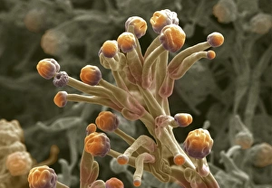
Background imageFungal Collection: Mould sporangia

Mould sporangia. Coloured scanning electron micrograph (SEM) of the sporangia (orange) of an unidentified mould fungus. The sporangia are the organs in which the asexual spores are produced

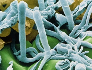
Background imageFungal Collection: Hyphae and asci of Sphaerotheca mildew

Hyphae and asci of Sphaerotheca mildew
Mildew. Coloured Scanning Electron Micrograph of hyphae and ripe asci of the leaf mildew Sphaerotheca sp. The hyphae are the tangled filaments which make up a fungal colony

Background imageFungal Collection: Yeast, Saccharomyces cerevisiae

Yeast, Saccharomyces cerevisiae
Yeast cells. Coloured scanning electron micro- graph (SEM) of budding yeast cells (yellow, Saccharomyces cerevisiae). Known as bakers or brewers yeast

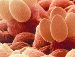
Background imageFungal Collection: Field Mushroom basidiospores

Field Mushroom basidiospores
Mushroom basidiospores. Coloured scanning electron micrograph (SEM) of basidiospores of the Field Mushroom (Agaricus campestris)

Background imageFungal Collection: Fungal growth on wet floorboards in derelict farmhouse - Cotswolds UK

Fungal growth on wet floorboards in derelict farmhouse - Cotswolds UK
MAB-1451 Fungal growth on wet floorboards in derelict farmhouse Cotswolds UK Mark Boulton contact details: prints@ardea.com tel: +44 (0) 20 8318 1401

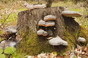
Background imageFungal Collection: Hoof fungus - on stump - Georgia

Hoof fungus - on stump - Georgia
ROG-13970 Hoof fungus - on stump Georgia. Fomitopsis pinicola Bob Gibbons contact details: prints@ardea.com tel: +44 (0) 20 8318 1401

Background imageFungal Collection: Cultivated Crabapple (Malus sp. ) close-up of leaves, infected with Cedar-apple Rust

Cultivated Crabapple (Malus sp. ) close-up of leaves, infected with Cedar-apple Rust
Cultivated Crabapple (Malus sp.) close-up of leaves, infected with Cedar-apple Rust (Gymnosporangium juniperi-virginianae), U.S.A

Background imageFungal Collection: Lemon (Citrus limon) close-up of fruit, damaged by Stem End Rot (Phomopsis citri)

Lemon (Citrus limon) close-up of fruit, damaged by Stem End Rot (Phomopsis citri)

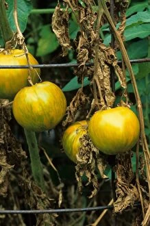
Background imageFungal Collection: Tomato (Solanum sp. ) Green Zebra, close-up of fruit, with Fusarium Wilt

Tomato (Solanum sp. ) Green Zebra, close-up of fruit, with Fusarium Wilt (Fusarium oxysporum lycopersici)
Tomato (Solanum sp.) Green Zebra, close-up of fruit, with Fusarium Wilt (Fusarium oxysporum lycopersici) causes vascular wilt of leaves, U.S.A

Background imageFungal Collection: Maize (Zea mays) crop, leaves damaged by Northern Leaf Blight (Exserohilum turcicum), Gisborne

Maize (Zea mays) crop, leaves damaged by Northern Leaf Blight (Exserohilum turcicum), Gisborne, North Island, New Zealand

Background imageFungal Collection: Grasshopper (Orthoptera sp. ) adult, killed by Pathogenic Fungus (Entomophthora sp)

Grasshopper (Orthoptera sp. ) adult, killed by Pathogenic Fungus (Entomophthora sp)
Grasshopper (Orthoptera sp.) adult, killed by Pathogenic Fungus (Entomophthora sp.) infection, fungus causes grasshopper to climb stalk before dying so spores will spread further, Pyrenees, Ariege

Background imageFungal Collection: Marsh Frog (Pelophylax ridibundus) dead adult, killed by fungal disease, at edge of pond habitat

Marsh Frog (Pelophylax ridibundus) dead adult, killed by fungal disease, at edge of pond habitat, Italy, april

Background imageFungal Collection: Agile Frog (Rana dalmatina) immature, close-up of underside showing fungal disease and infections

Agile Frog (Rana dalmatina) immature, close-up of underside showing fungal disease and infections, Italy, may

Background imageFungal Collection: Sycamore Tar Spot (Rhytisma acerinum) lesions on fallen Sycamore (Acer pseudoplatanus) leaves

Sycamore Tar Spot (Rhytisma acerinum) lesions on fallen Sycamore (Acer pseudoplatanus) leaves, covered with light snow, Romania, october

Background imageFungal Collection: Dance Fly (Empis tessellata) adult, fallen victim to Pathogenic Fungus (Entomophthora muscae)

Dance Fly (Empis tessellata) adult, fallen victim to Pathogenic Fungus (Entomophthora muscae) that enters brain and causes it to land, climb to the top of bush and die

Background imageFungal Collection: Black slug on waxcap fungus

Black slug on waxcap fungus
Black slug (Arion ater) on a splendid waxcap fungus (Hygrocybe splendidissima). The splendid waxcap is inedible to humans

Background imageFungal Collection: Wood wasp, SEM

Wood wasp, SEM
Wood wasp, coloured scanning electron micrograph (SEM). This wasp, also called a horntail, is in the family Siricidae. Females lay eggs in bark

Background imageFungal Collection: Wood wasp, SEM

Wood wasp, SEM
Wood wasp, coloured scanning electron micrograph (SEM). This wasp, also called a horntail, is in the family Siricidae. Females lay eggs in bark

Background imageFungal Collection: Phagocytosis of a yeast spore, SEM

Phagocytosis of a yeast spore, SEM
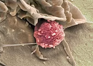
Phagocytosis of a yeast spore. Coloured scanning electron micrograph (SEM) of a neutrophil (brown) phagocytosing (engulfing and destroying) a spore from the yeast Candida albicans (orange)

Background imageFungal Collection: Phagocytosis of a fungus spore, SEM

Phagocytosis of a fungus spore, SEM
Phagocytosis of a fungus spore. Coloured scanning electron micrograph (SEM) of a spore from the fungus Aspergillus fumigatus (red) being phagocytosed (engulfed and destroyed)

Background imageFungal Collection: Fungal infections

Fungal infections, Artwork. Colonies of fungi on a petri dish with examples of fungal skin infections. In the background are fungal hyphae (filaments)

Background imageFungal Collection: Roquefort cheese

Roquefort cheese on a plate. Roquefort cheese is a French cheese originating from the Roquefort-sur- Soulzon region. It is made from cows milk

Background imageFungal Collection: Medical forceps, 18th century

Medical forceps, 18th century

Background imageFungal Collection: Fungal bladder infection, 18th century

Fungal bladder infection, 18th century

Background imageFungal Collection: Petri dish cultures

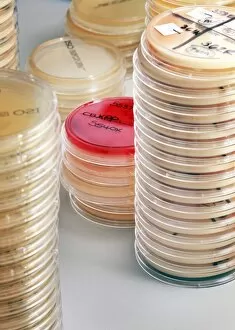
Petri dish cultures
Stacked petri dishes containing agar on which to culture cells, bacteria or fungi in a laboratory

Background imageFungal Collection: Shiitake mushroom

Shiitake mushroom (Lentinula edodes). Also known as black forest mushrooms, this fungus has been cultivated in the eastern Asia for over 650 years

Background imageFungal Collection: Penicillium mould colony

Penicillium mould colony. Penicillium is used to produce the antibiotic penicillin. Magnification: x60 when printed 10 centimetres wide

Background imageFungal Collection: Pear rust fungus, light micrograph

Pear rust fungus, light micrograph
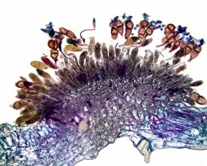
Pear rust fungus. Light micrograph of a section through tissue from a juniper tree infected with pear rust fungus (Gymnosporangium sabinae, lower right)

Background imageFungal Collection: Grey mould fungus, light micrograph

Grey mould fungus, light micrograph
Grey mould fungus. Light micrograph of a section through onion tissue infected with grey mould fungus (Botrytis allii). Botrytis is a parasitic saprophyte mould that grows on wounded plants

Background imageFungal Collection: Corn smut infection, light micrograph

Corn smut infection, light micrograph
Corn smut infection. Light micrograph of a section through tissue from a corn (Zea mays) plant infected with corn smut fungus (Ustilago maydis)

Background imageFungal Collection: Bramble rust fungus spores

Bramble rust fungus spores. Light micrograph of teliospores from the bramble rust fungus (Phragmidium violaceum). Magnification: x37 when printed 10 centimetres wide

Background imageFungal Collection: Rose mildew fungus, light micrograph

Rose mildew fungus, light micrograph
Rose mildew fungus. Light micrograph of a section through tissue from a rose plant infected with rose mildew fungus (Erysiphe pannosa)

Background imageFungal Collection: Pear scab infection, light micrograph

Pear scab infection, light micrograph
Pear scab infection. Light micrograph of a section through a pear (Pyrus sp.) infected with the Venturia pirinum fungus. This fungus causes scab, or black spot, of the pear fruit

Background imageFungal Collection: Black scab infection, light micrograph

Black scab infection, light micrograph
Black scab infection. Light micrograph of a section through a potato infected with black scab (Synchytrium endobioticum) fungus

Background imageFungal Collection: White rust fungus on a leaf

White rust fungus on a leaf. Light micrograph of a section through a plant leaf infected with white rust fungus (Albugo candida)

Background imageFungal Collection: Earth ball fungus, light micrograph

Earth ball fungus, light micrograph
Earth ball fungus. Light micrograph of a section through tissue from the fruiting body of an earth ball (Scleroderma vulgare) mushroom

Background imageFungal Collection: Plum pocket infection, light micrograph

Plum pocket infection, light micrograph
Plum pocket infection. Light micrograph of a section through a plum gall caused by the plum pocket fungus (Taphrina pruni)

Background imageFungal Collection: Rust fungus in a leaf, light micrograph

Rust fungus in a leaf, light micrograph
Rust fungus in a leaf. Light micrograph of a section through a wheat leaf infected with the parasitic Puccinia graminis rust

Background imageFungal Collection: Fly agaric fungus cap

Fly agaric fungus cap
Close up of fly agaric toadstool cap showing remnants of the protective veil. Berkshire, UK September 2011

Background imageFungal Collection: Penicillium mould, light micrograph

Penicillium mould, light micrograph
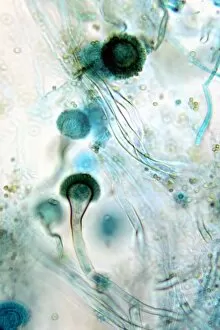
Penicillium mould. Light micrograph of a section through an orange rind infected with Penicillium sp. blue mould fungus, showing fungal hyphae and conidiophores (pink and purple) with spores (red)

Background imageFungal Collection: Brown mould fungus, light micrograph

Brown mould fungus, light micrograph
Brown mould fungus. Light micrograph of a section through decaying plant tissue infected with brown mould fungus (Aspergillus glaucum)

Background imageFungal Collection: Boletus fungus, light micrograph

Boletus fungus, light micrograph
Boletus fungus. Light micrograph of a section through tissue from the fruiting body of a Boletus sp. mushroom, showing the hymenium (dark red) of the fruiting body

Background imageFungal Collection: Hollyhock rust fungus in a leaf

Hollyhock rust fungus in a leaf. Light micrograph of a section through a hollyhock (Alcea sp.) leaf infected with the parasitic Puccinia malvacearum rust, showing the teliospores (spores)

Background imageFungal Collection: Yeast cell, artwork

Yeast cell, artwork
Yeast cell. Computer artwork showing the structure of a yeast cell


All Professionally Made to Order for Quick Shipping

"Fungal Kingdom: Exploring the Intricate World of Fungi" Budding yeast cell: Witness the remarkable process of reproduction as a budding yeast cell emerges, ready to create new life. Fly agaric mushrooms: Delve into the enchanting realm of fly agaric mushrooms, with their vibrant red caps and white spots, captivating both nature enthusiasts and fairytale lovers alike. SEM of penicillin fungus: Uncover the microscopic beauty of penicillin fungus through scanning electron microscopy (SEM), revealing its intricate structures that have revolutionized medicine. Culture of Aspergillus nidulans fungus: Step into a laboratory where cultures of Aspergillus nidulans fungus thrive, showcasing their unique growth patterns and potential for scientific research. Cep mushroom (Boletus edulis): Discover the culinary delight known as cep mushroom or Boletus edulis, renowned for its rich flavor and sought-after by chefs around the world. Candida fungus, SEM: Explore the unseen world within our bodies as Candida fungi are magnified under an electron microscope, shedding light on their role in various infections and diseases. Mushroom gills, SEM: Marvel at the intricate network of gills found beneath mushroom caps when observed through scanning electron microscopy (SEM), highlighting their crucial role in spore production. Fly agaric fungi: Journey through enchanted forests where fly agaric fungi flourish; these iconic red-and-white toadstools evoke a sense of wonderment while harboring fascinating ecological relationships with other organisms. Dividing yeast cells, SEM: Peer into a microscopic universe where dividing yeast cells undergo mitosis under high-resolution imaging techniques—witnessing life's continuous cycle unfold before your eyes. Penicillium roqueforti fungus: Meet Penicillium roqueforti—a blue mold used in cheese-making—whose distinctive flavor and appearance contribute to the creation of beloved blue cheeses worldwide.