Fraud Blocker Skip to main content

Fungal Collection (page 3)

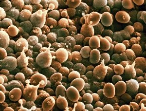
"Fungal Kingdom: Exploring the Intricate World of Fungi" Budding yeast cell: Witness the remarkable process of reproduction as a budding yeast cell emerges

Background imageFungal Collection: Box blight, Cylindrocladiumn buxicola, damage to Buxus sempervirens parterre hedge

Box blight, Cylindrocladiumn buxicola, damage to Buxus sempervirens parterre hedge

Background imageFungal Collection: Box rust, Puccia buxi, pustules on the lower surface of a diseased parterre hedge leaves

Box rust, Puccia buxi, pustules on the lower surface of a diseased parterre hedge leaves

Background imageFungal Collection: Branching threads of fungus mycelium in organic soil

Branching threads of fungus mycelium in organic soil

Background imageFungal Collection: Box rust, Puccia buxi, pustules on the upper surface of a diseased parterre hedge leaves

Box rust, Puccia buxi, pustules on the upper surface of a diseased parterre hedge leaves

Background imageFungal Collection: Brown-rot (Postia placenta) growing over decaying tree stump, Clumber Park, Nottinghamshire

Brown-rot (Postia placenta) growing over decaying tree stump, Clumber Park, Nottinghamshire, England, October

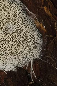
Background imageFungal Collection: Brown-rot (Postia placenta) close-up detail of pores, Clumber Park, Nottinghamshire, England

Brown-rot (Postia placenta) close-up detail of pores, Clumber Park, Nottinghamshire, England, October

Background imageFungal Collection: Fungus (Trechispora hymenocystis) spreading over dead wood with fungal hyphae around margins

Fungus (Trechispora hymenocystis) spreading over dead wood with fungal hyphae around margins, Clumber Park, Nottinghamshire, England, October

Background imageFungal Collection: Jelly Rot Fungus (Phlebia tremellosa) fruiting body, growing on decaying wood, Clumber Park

Jelly Rot Fungus (Phlebia tremellosa) fruiting body, growing on decaying wood, Clumber Park, Nottinghamshire, England, September

Background imageFungal Collection: Fungus (Taphrina sp. ) gall, abnormal outgrowth on underside of diseased leaf, Clumber Park

Fungus (Taphrina sp. ) gall, abnormal outgrowth on underside of diseased leaf, Clumber Park, Nottinghamshire, England
Fungus (Taphrina sp.) gall, abnormal outgrowth on underside of diseased leaf, Clumber Park, Nottinghamshire, England, October

Background imageFungal Collection: Scarlet Caterpillar Fungus (Cordyceps militaris) fruiting bodies, growing in woodland, Powys

Scarlet Caterpillar Fungus (Cordyceps militaris) fruiting bodies, growing in woodland, Powys, Wales, November

Background imageFungal Collection: Cultivated Apple (Malus domestica) McIntosh, close-up of leaf underside

Cultivated Apple (Malus domestica) McIntosh, close-up of leaf underside, with Frogeye (Sphaeropsis sp)
Cultivated Apple (Malus domestica) McIntosh, close-up of leaf underside, with Frogeye (Sphaeropsis sp.) Leaf Spot with spores visible, Ottawa, Ontario, Canada, August

Background imageFungal Collection: Freckled dapperling (Lepiota aspera) C018 / 1771

Freckled dapperling (Lepiota aspera) C018 / 1771
Freckled dapperling (Lepiota aspera) in a woodland clearing

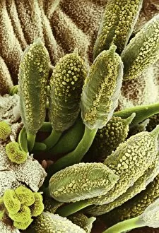
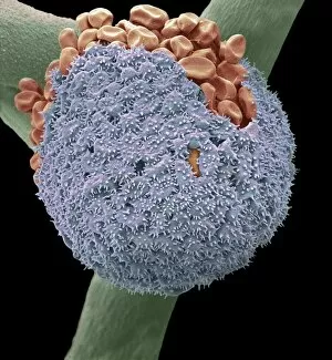
Background imageFungal Collection: Rust fungus on a rose leaf, SEM C017 / 7132

Rust fungus on a rose leaf, SEM C017 / 7132
Rust fungus on a rose leaf. Coloured scanning electron micrograph (SEM) of rust fungus (Phragmidium sp.) spores emerging from a rose (Rosa sp.) leaf (brown)

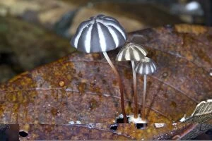
Background imageFungal Collection: Tropical mushrooms C014 / 0952

Tropical mushrooms C014 / 0952
Tropical mushrooms growing in leaf litter. Photographed in Rio Bigal Biological Reserve, Amazon Rainforest, Ecuador

Background imageFungal Collection: Fungal leaf spot C014 / 2095

Fungal leaf spot C014 / 2095
Fungal leaf spot/blotch on Acer davidii

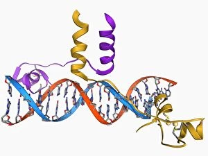
Background imageFungal Collection: Pho4 transcription factor bound to DNA C014 / 0861

Pho4 transcription factor bound to DNA C014 / 0861
Pho4 transcription factor bound to DNA. Molecular model showing phosphate system positive regulatory protein (Pho4) (blue and green) bound to a strand of DNA (deoxyribonucleic acid, red and purple)

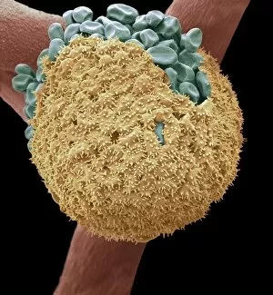
Background imageFungal Collection: Fungal spores on pollen grain, SEM

Fungal spores on pollen grain, SEM
Fungal spores on pollen grain. Coloured scanning electron micrograph (SEM) showing fungal spores (round) on a pollen grain from a goji berry (wolfberry, Lycium sp.) plant

Background imageFungal Collection: Bread mould, SEM C017 / 7127

Bread mould, SEM C017 / 7127
Bread mould. Coloured scanning electron micrograph (SEM) of a sporangium of a bread mould (Mucor sp.). Sporangia are asexual reproductive structures where the moulds spores develop

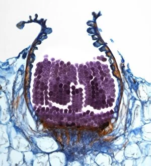
Background imageFungal Collection: Rust fungus spore, light micrograph F006 / 9817

Rust fungus spore, light micrograph F006 / 9817
Rust fungus spore. Light micrograph of a section through an aeciospore from a Puccinia sp. rust fungus. Spores are the reproductive structures of a fungus
Background imageFungal Collection: Bread mould, SEM C017 / 7129

Bread mould, SEM C017 / 7129
Bread mould. Coloured scanning electron micrograph (SEM) of a sporangium of a bread mould (Mucor sp.). Sporangia are asexual reproductive structures where the moulds spores develop

Background imageFungal Collection: Sir3 gene silencer acting on DNA F006 / 9730

Sir3 gene silencer acting on DNA F006 / 9730
Sir3 gene silencer acting on DNA, molecular model. Sir3 (light blue) is acting on a circular strand of DNA (deoxyribonucleic acid, pink)

Background imageFungal Collection: Fungal prion protein F006 / 9644

Fungal prion protein F006 / 9644
Fungal prion protein. Molecular model of the amyloid form of the HET prion protein

Background imageFungal Collection: Mitochondrial processing peptidase F006 / 9362

Mitochondrial processing peptidase F006 / 9362
Yeast mitochondrial processing peptidase, molecular model. This enzyme plays an essential role in importing mitochondrial proteins

Background imageFungal Collection: Yeast DNA recognition, molecular model F006 / 9282

Yeast DNA recognition, molecular model F006 / 9282
Yeast DNA recognition. Computer model showing a GAL4 transcription activator protein bound to a yeast DNA (deoxyribonucleic acid) molecule (red and blue)

Background imageFungal Collection: Cup lichen (Cladonia sp. )

Cup lichen (Cladonia sp. )
Cup lichen (Cladonia sp.) growing on a stone. Lichens are symbiotic organisms formed of a fungus and an alga that mutually benefit one another. Photographed in the UK

Background imageFungal Collection: Lichen, SEM C015 / 5082

Lichen, SEM C015 / 5082
Lichen, coloured environmental scanning electron micrograph (ESEM). Lichens are symbiotic organisms formed of a fungus and an alga that mutually benefit one another

Background imageFungal Collection: Pine cone on lichen

Pine cone on lichen. Close-up of a cone from a pine (Pinus sp.) tree on reindeer lichen (Cladonia portentosa). Photographed in the UK

Background imageFungal Collection: Black chanterelle mushrooms

Black chanterelle mushrooms (Craterellus cornucopioides). Photographed in the New Forest, Hampshire, UK

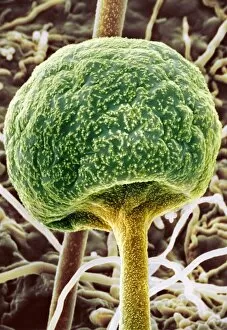
Background imageFungal Collection: Bread mould, SEM C016 / 9051

Bread mould, SEM C016 / 9051
Bread mould. Coloured scanning electron micrograph (SEM) of a black bread mould (Rhizopus stolonifer) sporangium (orange, spherical)

Background imageFungal Collection: Curvularia geniculata fungus, SEM C016 / 9389

Curvularia geniculata fungus, SEM C016 / 9389
Curvularia geniculata fungus. Coloured scanning electron micrograph (SEM) of a colony of the filamentous fungus Curvularia geniculata. This hyphomycete fungus is a pathogen of many plant species

Background imageFungal Collection: Apple tree fungus, SEM C016 / 9416

Apple tree fungus, SEM C016 / 9416
Apple tree fungus. Coloured scanning electron micrograph (SEM) of fungal spores bursting through a lesion on the surface of a leaf from an apple (Malus domestica) tree

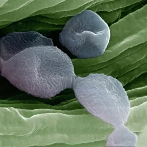
Background imageFungal Collection: Mould spores, SEM C016 / 9419

Mould spores, SEM C016 / 9419
Mould spores. Coloured scanning electron micrograph (SEM) of mould (fungal) spores (blue) on food (green)

Background imageFungal Collection: Bread mould, SEM C016 / 9052

Bread mould, SEM C016 / 9052
Bread mould. Coloured scanning electron micrograph (SEM) of a black bread mould (Rhizopus stolonifer) sporangium (blue, spherical)

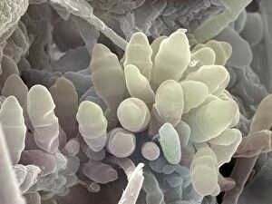
Background imageFungal Collection: Mushrooms spores, SEM C016 / 9450

Mushrooms spores, SEM C016 / 9450
Mushrooms spores. Coloured scanning electron micrograph (SEM) of budding spores in the basidium of an Agaricus bisporus fungus

Background imageFungal Collection: Fungus on orchid C016 / 7748

Fungus on orchid C016 / 7748
Fungus on orchid. Close-up of a tropical fungus growing on the leaves of an orchid (family Orchidaceae). Photographed in Yasuni National Park, Amazon rainforest, Ecuador

Background imageFungal Collection: Mould spores, SEM C016 / 9418

Mould spores, SEM C016 / 9418
Mould spores. Coloured scanning electron micrograph (SEM) of mould (fungal) spores (blue) on food (green)

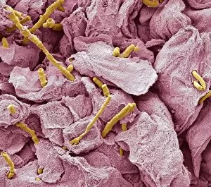
Background imageFungal Collection: Thrush infection of the tongue, SEM C016 / 9091

Thrush infection of the tongue, SEM C016 / 9091
Thrush infection of the tongue. Coloured scanning electron micrograph (SEM) of the surface of a tongue infected with Candida sp. fungus (yellow), known as yeast infection, thrush, or candidiasis

Background imageFungal Collection: Apple tree fungus, SEM C016 / 9417

Apple tree fungus, SEM C016 / 9417
Apple tree fungus. Coloured scanning electron micrograph (SEM) of fungal spores bursting through a lesion on the surface of a leaf from an apple (Malus domestica) tree

Background imageFungal Collection: Bread mould, SEM C016 / 9050

Bread mould, SEM C016 / 9050
Bread mould. Coloured scanning electron micrograph (SEM) of a black bread mould (Rhizopus stolonifer) sporangium (green, spherical)

Background imageFungal Collection: Soil structure and fauna, artwork

Soil structure and fauna, artwork
Soil structure and fauna. Artwork of a vertical section through soil, showing various structures and fauna. The surface layer includes organic mulch, the bottom layer is rocks

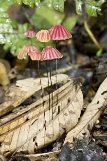
Background imageFungal Collection: Tropical mushrooms (Marasmius sp. ) C016 / 6240

Tropical mushrooms (Marasmius sp. ) C016 / 6240
Tropical mushrooms. Close-up of an agaric (Marasmius sp.) fungus growing in the leaf litter of the forest floor. Photographed in Yasuni National Park, Amazon rainforest, Ecuador

Background imageFungal Collection: Tropical mushrooms C016 / 6248

Tropical mushrooms C016 / 6248
Tropical mushrooms. Close-up of a fungus growing on a rotting log on the forest floor. Photographed in Yasuni National Park, Amazon rainforest, Ecuador

Background imageFungal Collection: Ant on a toadstool C016 / 6249

Ant on a toadstool C016 / 6249
Ant on a toadstool. Bulldog ant (Myrmecia sp.) on a Marasimus sp. toadstool on the rainforest floor. Photographed in Yasuni National Park, Amazon rainforest, Ecuador

Background imageFungal Collection: Bioluminescent mushrooms C016 / 6238

Bioluminescent mushrooms C016 / 6238
Bioluminescent mushrooms. Close-up of the bioluminescing fruiting bodies (mushrooms) of a tropical fungus growing on a rotting branch on the floor of a rainforest

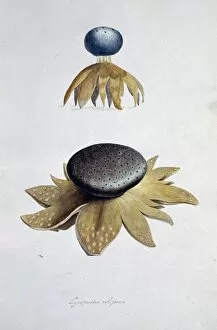
Background imageFungal Collection: Fungus (Lycoperdon coliforme) C016 / 5988

Fungus (Lycoperdon coliforme) C016 / 5988
Fungus (Lycoperdon coliforme). Plate 1 from Drawings of Submerged Algae (1800) by Mary Dawson Turner. Held in the Botany Library at the Natural History Museum, London, UK

Background imageFungal Collection: Mushroom, SEM C015 / 9162

Mushroom, SEM C015 / 9162
Mushroom, coloured scanning electron micrograph (SEM). Gills are seen on the underside of the mushrooms cap. These are the structures from which reproductive spores are shed

Background imageFungal Collection: Mushroom, SEM C015 / 9161

Mushroom, SEM C015 / 9161
Mushroom, coloured scanning electron micrograph (SEM). Gills are seen on the underside of the mushrooms cap. These are the structures from which reproductive spores are shed

Background imageFungal Collection: Rust fungus on a rose leaf, SEM C017 / 7131

Rust fungus on a rose leaf, SEM C017 / 7131
Rust fungus on a rose leaf. Coloured scanning electron micrograph (SEM) of rust fungus (Phragmidium sp.) spores emerging from a rose (Rosa sp.) leaf (brown)


All Professionally Made to Order for Quick Shipping

"Fungal Kingdom: Exploring the Intricate World of Fungi" Budding yeast cell: Witness the remarkable process of reproduction as a budding yeast cell emerges, ready to create new life. Fly agaric mushrooms: Delve into the enchanting realm of fly agaric mushrooms, with their vibrant red caps and white spots, captivating both nature enthusiasts and fairytale lovers alike. SEM of penicillin fungus: Uncover the microscopic beauty of penicillin fungus through scanning electron microscopy (SEM), revealing its intricate structures that have revolutionized medicine. Culture of Aspergillus nidulans fungus: Step into a laboratory where cultures of Aspergillus nidulans fungus thrive, showcasing their unique growth patterns and potential for scientific research. Cep mushroom (Boletus edulis): Discover the culinary delight known as cep mushroom or Boletus edulis, renowned for its rich flavor and sought-after by chefs around the world. Candida fungus, SEM: Explore the unseen world within our bodies as Candida fungi are magnified under an electron microscope, shedding light on their role in various infections and diseases. Mushroom gills, SEM: Marvel at the intricate network of gills found beneath mushroom caps when observed through scanning electron microscopy (SEM), highlighting their crucial role in spore production. Fly agaric fungi: Journey through enchanted forests where fly agaric fungi flourish; these iconic red-and-white toadstools evoke a sense of wonderment while harboring fascinating ecological relationships with other organisms. Dividing yeast cells, SEM: Peer into a microscopic universe where dividing yeast cells undergo mitosis under high-resolution imaging techniques—witnessing life's continuous cycle unfold before your eyes. Penicillium roqueforti fungus: Meet Penicillium roqueforti—a blue mold used in cheese-making—whose distinctive flavor and appearance contribute to the creation of beloved blue cheeses worldwide.