Fraud Blocker Skip to main content

Fungal Collection (page 5)

"Fungal Kingdom: Exploring the Intricate World of Fungi" Budding yeast cell: Witness the remarkable process of reproduction as a budding yeast cell emerges

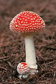
Background imageFungal Collection: Fly agaric (Amanita muscaria) fungus C014 / 3019

Fly agaric (Amanita muscaria) fungus C014 / 3019
Fly agaric (Amanita muscaria) fungus. These poisonous mushrooms are the fruiting bodies of the Amanita muscaria fungus. The caps have long been dried

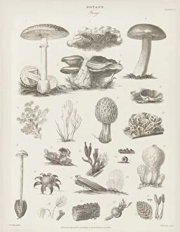
Background imageFungal Collection: Fungal fruiting bodies, 19th century C015 / 6087

Fungal fruiting bodies, 19th century C015 / 6087
Fungal fruiting bodies, 19th-century artwork. The shape of fungal fruiting bodies can range from capped and gilled toadstools and mushrooms (top), to bracket fungi (upper centre), morels (centre)

Background imageFungal Collection: Heat shock transcription factor and DNA C015 / 5558

Heat shock transcription factor and DNA C015 / 5558
Heat shock transcription factor and DNA. Molecular model of the binding domain region (purple) of a heat shock protein transcription factor bound to DNA (pink, deoxyribonucleic acid)

Background imageFungal Collection: Mycena epipterygia mushroom C018 / 2535

Mycena epipterygia mushroom C018 / 2535
Mycena epipterygia mushroom

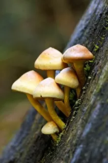
Background imageFungal Collection: Sulphur tuft (Hypholoma fasciculare) C018 / 2565

Sulphur tuft (Hypholoma fasciculare) C018 / 2565
Sulphur tuft (Hypholoma fasciculare) mushrooms

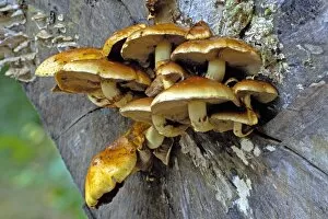
Background imageFungal Collection: Sulphur tuft (Hypholoma fasciculare) C018 / 2534

Sulphur tuft (Hypholoma fasciculare) C018 / 2534
Sulphur tuft (Hypholoma fasciculare) mushrooms

Background imageFungal Collection: Scleroderma citrinum mushroom C018 / 2538

Scleroderma citrinum mushroom C018 / 2538
Common earthball (Scleroderma citrinum) mushroom

Background imageFungal Collection: Ash dieback disease fungus, SEM C017 / 8028

Ash dieback disease fungus, SEM C017 / 8028
Ash dieback disease fungus. Coloured scanning electron micrograph (SEM) of the surface of a fruiting body of the fungus Chalara fraxinea

Background imageFungal Collection: Ash dieback disease fungus, SEM C017 / 8026

Ash dieback disease fungus, SEM C017 / 8026
Ash dieback disease fungus. Coloured scanning electron micrograph (SEM) of a fruiting body of the fungus Chalara fraxinea, also known as Hymenoscyphus pseudoalbidus in its fruiting stage

Background imageFungal Collection: Ash dieback disease fungus, SEM C017 / 8027

Ash dieback disease fungus, SEM C017 / 8027
Ash dieback disease fungus. Coloured scanning electron micrograph (SEM) of fruiting bodies of the fungus Chalara fraxinea, also known as Hymenoscyphus pseudoalbidus in its fruiting stage

Background imageFungal Collection: Shaggy pholiota (Pholiota aurivella) C018 / 2566

Shaggy pholiota (Pholiota aurivella) C018 / 2566
Shaggy pholiota (Pholiota aurivella) mushrooms

Background imageFungal Collection: Beechwood sickener (Russula nobilis) C018 / 2532

Beechwood sickener (Russula nobilis) C018 / 2532
Beechwood sickener (Russula nobilis) mushroom

Background imageFungal Collection: Shaggy pholiota (Pholiota aurivella) C018 / 2533

Shaggy pholiota (Pholiota aurivella) C018 / 2533
Shaggy pholiota (Pholiota aurivella) mushrooms

Background imageFungal Collection: Fungus on a redwood tree C018 / 2574

Fungus on a redwood tree C018 / 2574
Fungus on a redwood tree. Tree fungus growing on a coast redwood (Sequoia sempervirens) tree. Photographed in Muir Woods National Park, California, USA
Background imageFungal Collection: Hypholoma mushrooms C018 / 2568

Hypholoma mushrooms C018 / 2568
Hypholoma mushrooms. Sulphur tuft (Hypholoma fasciculare, left) and brick cap (Hypholoma sublateritium, right) mushrooms growing on a tree stump

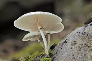
Background imageFungal Collection: Porcelain fungus (Oudemansiella mucida) C018 / 2564

Porcelain fungus (Oudemansiella mucida) C018 / 2564
Porcelain fungus (Oudemansiella mucida) mushroom

Background imageFungal Collection: Mycena epipterygia mushroom C018 / 2536

Mycena epipterygia mushroom C018 / 2536
Mycena epipterygia mushroom

Background imageFungal Collection: Early Devonian landscape, artwork C016 / 5348

Early Devonian landscape, artwork C016 / 5348
Early Devonian landscape. Artwork of early vascular plants (Rhynia) and larger fungal or lichen-like organisms (Prototaxites)

Background imageFungal Collection: Zombie ant fungus life-cycle, artwork

Zombie ant fungus life-cycle, artwork
Zombie ant fungus life-cycle. Computer artwork showing the life cycle of the parasitoidal fungus Ophiocordyceps unilateralis

Background imageFungal Collection: Oak Mazegill (Daedalea quercina) C016 / 3601

Oak Mazegill (Daedalea quercina) C016 / 3601
Oak Mazegill (Daedalea quercina) growing on an oak log in autumn. Photographed in Exmoor National Park, Devon

Background imageFungal Collection: Devils Fingers (Clathrus archeri)

Devils Fingers (Clathrus archeri), also known as Octopus stinkhorn. This is a fungus, introduced from Australia and naturalised in Europe

Background imageFungal Collection: Scutiger pes-caprae-sabatera mushrooms C016 / 3446

Scutiger pes-caprae-sabatera mushrooms C016 / 3446
Scutiger pes-caprae-sabatera mushrooms, artwork

Background imageFungal Collection: Caesars mushrooms (Amanita caesarea)

Caesars mushrooms (Amanita caesarea), artwork

Background imageFungal Collection: Puffball (Lycoperdon perlatum) mushrooms C016 / 3434

Puffball (Lycoperdon perlatum) mushrooms C016 / 3434
Puffball (Lycoperdon perlatum) mushrooms, artwork

Background imageFungal Collection: Fairy ring (Marasmius oreades) mushrooms C016 / 3433

Fairy ring (Marasmius oreades) mushrooms C016 / 3433
Fairy ring (Marasmius oreades) mushrooms, artwork

Background imageFungal Collection: Lactarius deliciosus mushrooms, artwork C016 / 3367

Lactarius deliciosus mushrooms, artwork C016 / 3367
Saffron milk cap (Lactarius deliciosus) mushrooms, artwork

Background imageFungal Collection: Hygrophorus russula mushrooms, artwork C016 / 3364

Hygrophorus russula mushrooms, artwork C016 / 3364
Hygrophorus russula mushrooms, artwork

Background imageFungal Collection: Oyster mushrooms on a tree C013 / 9918

Oyster mushrooms on a tree C013 / 9918
Oyster mushrooms (Pleurotus ostreatus) on a tree in woodland. These bracket mushrooms are edible fruiting bodies of the fungus, formed as it acts on dead and decaying wood

Background imageFungal Collection: Bare-root mycorrhizal fungi treatment C013 / 7276

Bare-root mycorrhizal fungi treatment C013 / 7276
Application of a mycorrhizal fungi product in the planting of a bare-root hedging plant. These fungi act as a plant stimulator by improving the access to nutrients

Background imageFungal Collection: Aspergillus fungus, artwork C013 / 4613

Aspergillus fungus, artwork C013 / 4613
Aspergillus fungus. Computer artwork of the fruiting body (conidiophore) of a typical mould (Aspergillus sp.), showing spores (conidia, round)

Background imageFungal Collection: Weeping widow mushrooms C013 / 5107

Weeping widow mushrooms C013 / 5107
Weeping widow mushrooms (Lacrymaria velutina). Photographed in autumn, in Normandy, France

Background imageFungal Collection: Giant puffball fungus in a basket C013 / 5108

Giant puffball fungus in a basket C013 / 5108
Giant puffball fungus (Calvatia gigantea) in a basket with a shallot and a bulb of garlic. Photographed in autumn, in Cornwall, UK

Background imageFungal Collection: Common ink cap mushrooms C013 / 5106

Common ink cap mushrooms C013 / 5106
Common ink cap mushrooms (Coprinopsis atramentaria). Photographed in autumn, in Normandy, France

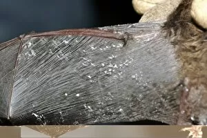
Background imageFungal Collection: White nose syndrome in bats C013 / 5105

White nose syndrome in bats C013 / 5105
White nose syndrome in bats. Wing of a little brown bat (Myotis lucifugus) with white nose syndrome (WNS). This deadly infection with the fungus Geomyces destructans leads to white fungal growths

Background imageFungal Collection: Tagged bat and white nose syndrome C013 / 5103

Tagged bat and white nose syndrome C013 / 5103
Tagged bat and white nose syndrome. Group of bats hibernating in a cave, one with an attached datalogger (yellow) and one with white nose syndrome (WNS)

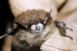
Background imageFungal Collection: White nose syndrome in bats C013 / 5104

White nose syndrome in bats C013 / 5104
White nose syndrome in bats. Nose of a little brown bat (Myotis lucifugus) with white nose syndrome (WNS). This deadly infection with the fungus Geomyces destructans leads to white fungal growths

Background imageFungal Collection: Silver birch bark (Betula pendula), SEM

Silver birch bark (Betula pendula), SEM
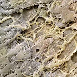
Detail of silver birch bark (Betula pendula), coloured scanning electron micrograph (SEM). Threads of fungal hyphae can be seen infiltrating the bark. Magnification x2000 at an image size of 10cm wide

Background imageFungal Collection: Mould fungus, SEM

Mould fungus, SEM
Mould fungus (Penicillium expansum), coloured scanning electron micrograph (SEM). This fungus, also called blue mould, is a pathogen of plant material

Background imageFungal Collection: Russula emetica fungus

Russula emetica fungus growing on moss. Known as the sickener mushroom, this poisonous fungus is usually found growing in woodland areas on the forest floor. Photographed in the UK

Background imageFungal Collection: Powdery mildew, SEM

Powdery mildew, SEM
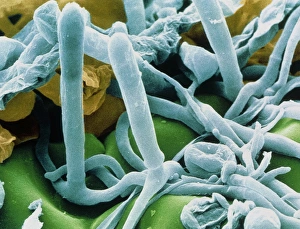
Powdery mildew. Coloured scanning electron micrograph (SEM) of the parasitic powdery mildew Erysiphe sp. on the stalk of a plant

Background imageFungal Collection: Mouldy crop, SEM

Mouldy crop, SEM
Mouldy crop. Coloured scanning electron micrograph (SEM) of grey rot (Botrytis sp.) on a runner bean plant. This is a parasitic fungus that can cause great damage to food crops

Background imageFungal Collection: Cabbage root infection, SEM

Cabbage root infection, SEM
Cabbage root infection. Coloured scanning electron micrograph (SEM) of a cross-section through one of the roots of a cabbage (Brassica sp)

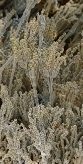
Background imageFungal Collection: Penicillium fungal spores, SEM

Penicillium fungal spores, SEM
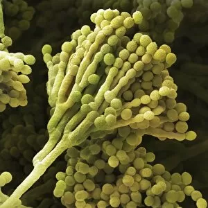
Penicillium fungal spores. Coloured scanning electron micrograph (SEM) of fruiting bodies of the fungus Penicillium roqueforti

Background imageFungal Collection: Penicillium spores

Penicillium spores
Penicillium. Coloured scanning electron micrograph (SEM) of the mycelium and fruiting bodies of the fungus Penicillium sp

Background imageFungal Collection: Fungus on a fly

Fungus on a fly
Fungus on fly. Coloured scanning electron micrograph (SEM) of an entomogenous fungus, Empusa muscae erupting from the abdomen of a housefly (Musca domestica)

Background imageFungal Collection: Mould sporangia

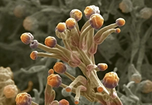
Mould sporangia. Coloured scanning electron micrograph (SEM) of the sporangia (orange) of an unidentified mould fungus. The sporangia are the organs in which the asexual spores are produced

Background imageFungal Collection: Hyphae and asci of Sphaerotheca mildew

Hyphae and asci of Sphaerotheca mildew
Mildew. Coloured Scanning Electron Micrograph of hyphae and ripe asci of the leaf mildew Sphaerotheca sp. The hyphae are the tangled filaments which make up a fungal colony

Background imageFungal Collection: Yeast, Saccharomyces cerevisiae

Yeast, Saccharomyces cerevisiae
Yeast cells. Coloured scanning electron micro- graph (SEM) of budding yeast cells (yellow, Saccharomyces cerevisiae). Known as bakers or brewers yeast


All Professionally Made to Order for Quick Shipping

"Fungal Kingdom: Exploring the Intricate World of Fungi" Budding yeast cell: Witness the remarkable process of reproduction as a budding yeast cell emerges, ready to create new life. Fly agaric mushrooms: Delve into the enchanting realm of fly agaric mushrooms, with their vibrant red caps and white spots, captivating both nature enthusiasts and fairytale lovers alike. SEM of penicillin fungus: Uncover the microscopic beauty of penicillin fungus through scanning electron microscopy (SEM), revealing its intricate structures that have revolutionized medicine. Culture of Aspergillus nidulans fungus: Step into a laboratory where cultures of Aspergillus nidulans fungus thrive, showcasing their unique growth patterns and potential for scientific research. Cep mushroom (Boletus edulis): Discover the culinary delight known as cep mushroom or Boletus edulis, renowned for its rich flavor and sought-after by chefs around the world. Candida fungus, SEM: Explore the unseen world within our bodies as Candida fungi are magnified under an electron microscope, shedding light on their role in various infections and diseases. Mushroom gills, SEM: Marvel at the intricate network of gills found beneath mushroom caps when observed through scanning electron microscopy (SEM), highlighting their crucial role in spore production. Fly agaric fungi: Journey through enchanted forests where fly agaric fungi flourish; these iconic red-and-white toadstools evoke a sense of wonderment while harboring fascinating ecological relationships with other organisms. Dividing yeast cells, SEM: Peer into a microscopic universe where dividing yeast cells undergo mitosis under high-resolution imaging techniques—witnessing life's continuous cycle unfold before your eyes. Penicillium roqueforti fungus: Meet Penicillium roqueforti—a blue mold used in cheese-making—whose distinctive flavor and appearance contribute to the creation of beloved blue cheeses worldwide.