Fraud Blocker Skip to main content

Diagnosis Collection (page 13)

"Unlocking the Secrets: The Power of Diagnosis" A seahorse's hidden world revealed through an X-ray, reminding us that diagnosis goes beyond human health

Background imageDiagnosis Collection: Pinned jaw fracture, X-ray C017 / 7982

Pinned jaw fracture, X-ray C017 / 7982
Pinned jaw fracture. X-ray of the head of a 22 year old male patient who has had a fractured mandible (lower jaw bone) pinned (lower left)

Background imageDiagnosis Collection: Duplex kidney collecting system, X-ray C017 / 7973

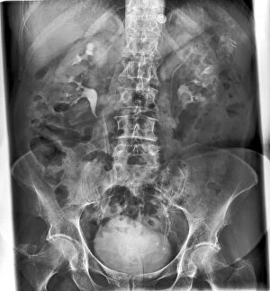
Duplex kidney collecting system, X-ray C017 / 7973
Duplex kidney collecting system. X-ray of the abdomen of a 78 year old male patient who has bilateral duplex collecting systems of the kidneys

Background imageDiagnosis Collection: Nail in foot, X-ray C017 / 7980

Nail in foot, X-ray C017 / 7980
Nail in foot. X-ray of the foot of a 7 year old female patient who had stood on a nail

Background imageDiagnosis Collection: Broken elbow, X-ray C017 / 7979

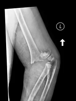
Broken elbow, X-ray C017 / 7979
Broken elbow. X-ray of the elbow of a 4 year old female with a displaced supracondylar fracture. The fracture is at the base of the humerus, the upper arm bone (upper frame)

Background imageDiagnosis Collection: Pinned broken ankle, X-ray C017 / 7978

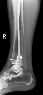
Pinned broken ankle, X-ray C017 / 7978
Pinned broken ankle. X-ray of the ankle of a 32 year old male that has been pinned after a parachuting accident. The ankle was dislocated between the tibia (shin bone)

Background imageDiagnosis Collection: Fractured and dislocated ankle, X-ray C017 / 7976

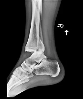
Fractured and dislocated ankle, X-ray C017 / 7976
Fractured and dislocated ankle. X-ray of the ankle of a 32 year old male who has been in a parachuting accident. The ankle is dislocated between the tibia (shin bone)

Background imageDiagnosis Collection: Infected malunited fracture, X-ray C017 / 7972

Infected malunited fracture, X-ray C017 / 7972
Infected malunited fracture. X-ray of the arm of a 65 year old female patient who has a bone infection after the nonunion of a fracture of the humerus (upper arm bone)

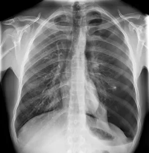
Background imageDiagnosis Collection: Pneumothorax, X-ray C017 / 7966

Pneumothorax, X-ray C017 / 7966
Pneumothorax. X-ray of the chest of a 22 year old male patient with a pneumothorax, or collapsed lung. The left lung (right on X-ray) has collapsed due to a build-up of air (solid grey)

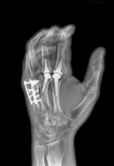
Background imageDiagnosis Collection: Knuckle replacement, X-ray C017 / 7971

Knuckle replacement, X-ray C017 / 7971
Knuckle replacement. X-ray of the hand of a 41 year old male patient who has had two of the joints replaced between the hand bones (metacarpals) and the first finger bones (proximal phalanges)

Background imageDiagnosis Collection: Broken elbow, X-ray C017 / 7968

Broken elbow, X-ray C017 / 7968
Broken elbow. X-ray of the elbow of a 5 year old female with a displaced supracondylar fracture. The fracture is at the base of the humerus, the upper arm bone (upper frame)

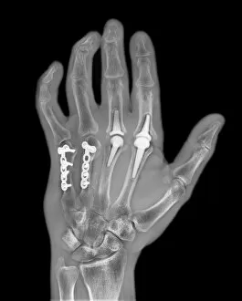
Background imageDiagnosis Collection: Knuckle replacement, X-ray C017 / 7970

Knuckle replacement, X-ray C017 / 7970
Knuckle replacement. X-ray of the hand of a 41 year old male patient who has had two of the joints replaced between the hand bones (metacarpals) and the first finger bones (proximal phalanges)

Background imageDiagnosis Collection: Pinned fractured elbow, X-ray C017 / 7969

Pinned fractured elbow, X-ray C017 / 7969
Pinned fractured elbow. X-ray of the elbow of a 5 year old female that has been internally fixed after a displaced supracondylar fracture

Background imageDiagnosis Collection: Reversed heart position, X-ray C017 / 7965

Reversed heart position, X-ray C017 / 7965
Reversed heart position. X-ray of the chest of a patient with dextrocardia, the congenital (present at birth) placement of the heart on the right side of the body

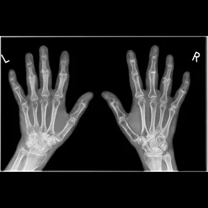
Background imageDiagnosis Collection: Arthritic hands, X-ray C017 / 7967

Arthritic hands, X-ray C017 / 7967
Arthritic hands. X-ray of the hands of a 49 year old female patient with arthritis of the finger joints. Arthritis is a degenerative disease that causes the loss of the protective cartilage between

Background imageDiagnosis Collection: Lung cancer, X-ray C017 / 7964

Lung cancer, X-ray C017 / 7964
Lung cancer. X-ray if the chest of a patient with malignant (cancerous) tumours in middle of the left lung (seen at right). The main cause of lung cancer is cigarette smoke

Background imageDiagnosis Collection: Knife in hand, X-ray C017 / 7963

Knife in hand, X-ray C017 / 7963
Knife in hand. X-ray of a knife through the left hand of a patient

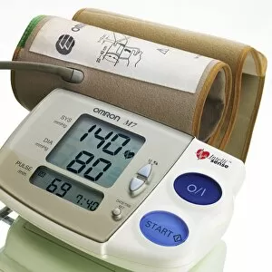
Background imageDiagnosis Collection: Digital blood pressure monitor C018 / 2880

Digital blood pressure monitor C018 / 2880
Digital blood pressure monitor (sphygmomanometer). This device is used to measure a persons blood pressure, an important indicator of health

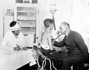
Background imageDiagnosis Collection: Tuberculosis campaign, early 20th century C017 / 7885

Tuberculosis campaign, early 20th century C017 / 7885
Tuberculosis campaign, early 20th century. Doctor using a stethoscope to check a boys lungs for signs of the respiratory bacterial disease tuberculosis (TB)

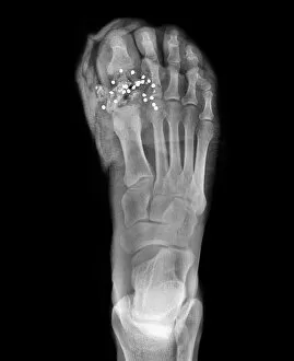
Background imageDiagnosis Collection: Firearm injury, X-ray C017 / 7873

Firearm injury, X-ray C017 / 7873
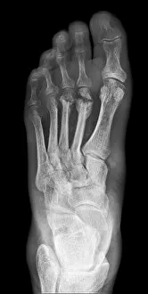
Firearm injury. X-ray of the foot of a patient who has been shot with a shotgun. Multiple pellets (white dots) can be seen in the big toe

Background imageDiagnosis Collection: Gallstones, X-ray C017 / 7860

Gallstones, X-ray C017 / 7860
Gallstones. X-ray of the abdomen of a patient showing four gallstones (white balls at upper left). Gallstones (choleliths)

Background imageDiagnosis Collection: Bone infection, X-ray C017 / 7861

Bone infection, X-ray C017 / 7861
Bone infection. X-ray of the foot of a diabetic patient osteomyelitis, a bacterial infection of the bone. Tissue damage to the foot caused by diabetes has lead to the infection

Background imageDiagnosis Collection: Ankle fracture with bone cyst, X-ray C017 / 7862

Ankle fracture with bone cyst, X-ray C017 / 7862
Ankle fracture. X-ray of the fractured ankle of a patient. The bone had been weakened by a cyst, a fluid-filled cavity

Background imageDiagnosis Collection: Large bowel obstruction, X-ray C017 / 7855

Large bowel obstruction, X-ray C017 / 7855
Large bowel obstruction. X-ray of the abdomen of a patient with an obstructed large intestine. The obstruction may be due to cancer, twisting of the intestine, an injury or an infection

Background imageDiagnosis Collection: Gallstone, Doppler ultrasound scan C017 / 7859

Gallstone, Doppler ultrasound scan C017 / 7859
Gallstone. Doppler ultrasound scan of the abdomen of a patient that has a gallstone in the common bile duct. Gallstones (choleliths)

Background imageDiagnosis Collection: Gallstone, ultrasound scan C017 / 7858

Gallstone, ultrasound scan C017 / 7858
Gallstone. Ultrasound scan of the abdomen of a patient that has a gallstone in the common bile duct. Gallstones (choleliths)

Background imageDiagnosis Collection: Large bowel obstruction, X-ray C017 / 7854

Large bowel obstruction, X-ray C017 / 7854
Large bowel obstruction. X-ray of the abdomen of a patient with an obstructed large intestine. The obstruction may be due to cancer, twisting of the intestine, an injury or an infection

Background imageDiagnosis Collection: Gallstone, ultrasound scan C017 / 7857

Gallstone, ultrasound scan C017 / 7857
Gallstone. Ultrasound scan of the abdomen of a patient that has a gallstone (white, lower centre) in the common bile duct

Background imageDiagnosis Collection: Kidney stones, X-ray C017 / 7856

Kidney stones, X-ray C017 / 7856
Kidney stones. X-ray of the kidney of a patient with kidney stones (dark areas) in the renal pelvis (upper centre) of the kidney

Background imageDiagnosis Collection: Pacemaker and heart pump, X-ray C017 / 7833

Pacemaker and heart pump, X-ray C017 / 7833
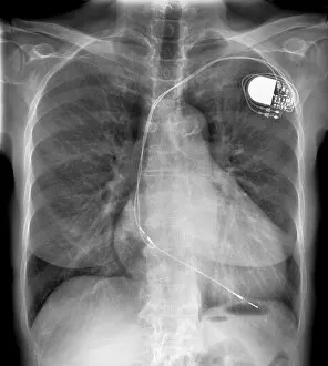
Pacemaker and heart pump. X-ray of the chest of a patient who is waiting for a heart and lung transplant. They have a pacemaker and a left ventricular assist device (LVAD) fitted

Background imageDiagnosis Collection: Diverticulitis C017 / 7835

Diverticulitis C017 / 7835
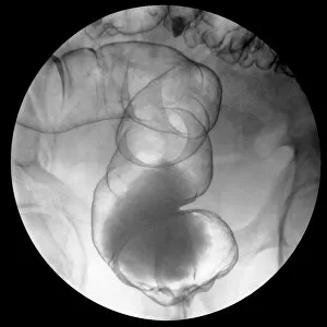
Diverticulitis. X-ray of the colon of a patient with diverticulitis. A diverticulum is a small pouch-like structure that protrudes out of weak spots in the intestinal wall

Background imageDiagnosis Collection: Enlarged heart, X-ray C017 / 7830

Enlarged heart, X-ray C017 / 7830
Enlarged heart. X-ray of the chest of a female patient with an enlarged heart. Cardiac diseases can make the heart need to pump harder

Background imageDiagnosis Collection: Enlarged heart, X-ray C017 / 7827

Enlarged heart, X-ray C017 / 7827
Enlarged heart. X-ray of the chest of a patient with an enlarged heart and a pacemaker (top right). Cardiac diseases can make the heart need to pump harder

Background imageDiagnosis Collection: Reversed heart position, X-ray C017 / 7825

Reversed heart position, X-ray C017 / 7825
Reversed heart position. X-ray of the chest of a patient with dextrocardia, the congenital (present at birth) placement of the heart on the right side of the body

Background imageDiagnosis Collection: Capsule endoscopy

Capsule endoscopy. Doctor holding a capsule endoscope. This pill-shaped capsule contains a small camera and is swallowed by a patient to provide real-time video images of the stomach and intestines

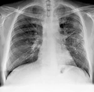
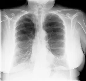
Background imageDiagnosis Collection: Lung disease, X-ray C017 / 7817

Lung disease, X-ray C017 / 7817
Lung disease. X-ray of the chest of a patient with chronic obstructive pulmonary disease (COPD). This is a collection of diseases that includes emphysema and chronic bronchitis

Background imageDiagnosis Collection: Pleural effusion, X-ray C017 / 7811

Pleural effusion, X-ray C017 / 7811
Pleural effusion. X-ray of the chest of a patient with pleural effusion of the left lung (seen on right). Pleural effusion is the accumulation of fluid in the spaces around the lungs

Background imageDiagnosis Collection: Pleural effusion, X-ray C017 / 7806

Pleural effusion, X-ray C017 / 7806
Pleural effusion. X-ray of the chest of a patient with bilateral pleural effusion. Pleural effusion is the accumulation of fluid in the spaces around the lungs

Background imageDiagnosis Collection: Dislocated hip replacement, X-ray C017 / 7804

Dislocated hip replacement, X-ray C017 / 7804
Dislocated hip replacement. X-ray of the pelvis of a patient whos total hip replacement of the rigt hip (left) has become dislocated

Background imageDiagnosis Collection: Ankle bone fracture, X-ray C017 / 7809

Ankle bone fracture, X-ray C017 / 7809
Ankle bone fracture. X-ray of the ankle of a patient with a fractured talus (ankle bone)

Background imageDiagnosis Collection: Dislocated hip replacement, X-ray C017 / 7803

Dislocated hip replacement, X-ray C017 / 7803
Dislocated hip replacement. X-ray of the pelvis of a patient whos total hip replacement of the rigt hip (left) has become dislocated

Background imageDiagnosis Collection: Ankle bone fracture, X-ray C017 / 7808

Ankle bone fracture, X-ray C017 / 7808
Ankle bone fracture. X-ray of the ankle of a patient with a fractured talus (ankle bone, lower centre)

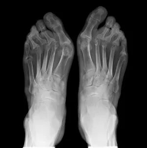
Background imageDiagnosis Collection: Arthritic feet, X-ray C017 / 7807

Arthritic feet, X-ray C017 / 7807
Arthritic feet. X-ray of the feet of a patient with rheumatoid arthritis. Rheumatoid arthritis is caused by the immune system attacking the bodys own tissues

Background imageDiagnosis Collection: Dislocated thumb, X-ray C017 / 7805

Dislocated thumb, X-ray C017 / 7805
Dislocated thumb. X-ray of the right hand of a 10 year old male patient with a dislocated thumb

Background imageDiagnosis Collection: Pulmonary embolism, X-ray C017 / 7802

Pulmonary embolism, X-ray C017 / 7802
Pulmonary embolism. X-ray of the chest of a 72 year old male patient with a pulmonary embolism. This is a blockage of the pulmonary artery

Background imageDiagnosis Collection: Endometrial polyp, ultrasound scan C017 / 7799

Endometrial polyp, ultrasound scan C017 / 7799
Endometrial polyp. Doppler ultrasound scan of the uterus (womb) of a 42 year old female patient with a polyp (lower centre) in the endometrium, the lining of the womb

Background imageDiagnosis Collection: Gallstone, ultrasound scan C017 / 7798

Gallstone, ultrasound scan C017 / 7798
Gallstone. Ultrasound scan of the gallbladder of a 81 year old male patient showing a gallstone measuring 5 centimetres in the neck of the gallbladder

Background imageDiagnosis Collection: Bladder mass, ultrasound scan C017 / 7796

Bladder mass, ultrasound scan C017 / 7796
Bladder mass. Ultrasound scan showing a large mass (centre) in the bladder of an 84 year old male patient. This could be a malignant (cancerous) tumour

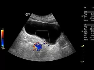
Background imageDiagnosis Collection: Bladder mass, Doppler ultrasound C017 / 7795

Bladder mass, Doppler ultrasound C017 / 7795
Bladder mass. Doppler ultrasound scan showing a large mass (black) with increased blood flow in the bladder of an 84 year old male patient. This could be a malignant (cancerous) tumour


All Professionally Made to Order for Quick Shipping

"Unlocking the Secrets: The Power of Diagnosis" A seahorse's hidden world revealed through an X-ray, reminding us that diagnosis goes beyond human health. Unveiling the spectrum: a color blindness test helping us understand how our eyes perceive the world differently. Eyesight test chart guiding us towards clarity and precision in diagnosing visual impairments. Peering into fractures: a broken wrist bone captured on X-ray, aiding doctors in accurate diagnosis and treatment plans. Exploring beneath the surface: a panoramic dental X-ray exposing hidden oral issues for comprehensive diagnoses. Empowering individuals with knowledge: home cholesterol test kits providing early detection for proactive health management. Saving lives through swift intervention: Pneumothorax treatment guided by an X-ray to restore lung function effectively. Knees tell stories too: a normal knee showcased on an X-ray, enabling precise diagnoses of joint conditions and injuries. Delving deeper into mysteries within: full body scans via MRI revealing intricate details for accurate medical assessments. Artistry meets science as ECGs portray the rhythm of a healthy heart, serving as diagnostic masterpieces in healthcare settings. Battling tuberculosis head-on with an X-ray, unveiling its presence so prompt treatments can be initiated promptly