Fraud Blocker Skip to main content

Diagnosis Collection (page 9)

"Unlocking the Secrets: The Power of Diagnosis" A seahorse's hidden world revealed through an X-ray, reminding us that diagnosis goes beyond human health

Background imageDiagnosis Collection: Normal skull, 3D CT scan C016 / 6326

Normal skull, 3D CT scan C016 / 6326
Normal skull. Coloured 3D computed tomography (CT) scan of the healthy skull of a 35 year old patient

Background imageDiagnosis Collection: Normal skull, 3D CT scan C016 / 6331

Normal skull, 3D CT scan C016 / 6331
Normal skull. Coloured cut-away 3D computed tomography (CT) scan of the healthy skull of a 35 year old patient

Background imageDiagnosis Collection: Human head, 3D CT scan C016 / 6393

Human head, 3D CT scan C016 / 6393
Human head. Coloured 3D computed tomography (CT) scan of a profile view of a 30 year old patients head and neck

Background imageDiagnosis Collection: Normal skull, 3D CT scan C016 / 6327

Normal skull, 3D CT scan C016 / 6327
Normal skull. Coloured 3D computed tomography (CT) scan of the healthy skull of a 35 year old patient

Background imageDiagnosis Collection: Human head, 3D CT scan C016 / 6377

Human head, 3D CT scan C016 / 6377
Human head. Coloured 3D computed tomography (CT) scan of a profile view of a 47 year old patients head. The brain is purple and bone is light brown. At centre, behind the face, are the sinuses

Background imageDiagnosis Collection: Bird flu diagnostic kit C015 / 8805

Bird flu diagnostic kit C015 / 8805
Bird flu diagnostic kit. Reagents for diagnosing influenza type a H7N9 infection in humans. This virus first emerged in the human population in China, in March 2013

Background imageDiagnosis Collection: Lung silicosis, light micrograph C015 / 7129

Lung silicosis, light micrograph C015 / 7129
Lung silicosis. Light micrograph of a section through a lung with silicosis, a type of pneumoconiosis caused by exposure to silica dusts. The lung tissue has become fibrous (centre)

Background imageDiagnosis Collection: Stethoscope demonstration C015 / 8835

Stethoscope demonstration C015 / 8835
MODEL RELEASED. Stethoscope demonstration. Medical worker showing how a stethoscope can be used to listen to your own heartbeat

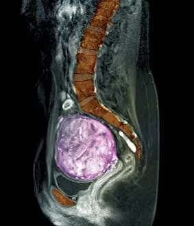
Background imageDiagnosis Collection: Uterine fibroids, MRI scan C018 / 0467

Uterine fibroids, MRI scan C018 / 0467
Uterine fibroids. Coloured magnetic resonance imaging (MRI) scan of the pelvis of a 47 year old female with two large fibroids (pink) in her uterus

Background imageDiagnosis Collection: Polycystic kidneys, CT scans C018 / 0571

Polycystic kidneys, CT scans C018 / 0571
Polycystic kidneys. Coloured computed tomography (CT) scans of the abdomen of a 28 year old patient with polycystic kidneys (pink)

Background imageDiagnosis Collection: Rectal cancer, MRI scans C018 / 0508

Rectal cancer, MRI scans C018 / 0508
Rectal cancer. Coloured magnetic resonance imaging (MRI) scans of the pelvis of a 65 year old patient with a malignant (cancerous) tumour (purple) in the rectum, the last part of the large intestine

Background imageDiagnosis Collection: Rectal cancer, MRI scans C018 / 0510

Rectal cancer, MRI scans C018 / 0510
Rectal cancer. Coloured magnetic resonance imaging (MRI) scans of the pelvis of a 65 year old patient with a malignant (cancerous) tumour in the rectum, the last part of the large intestine

Background imageDiagnosis Collection: Polycystic kidneys, CT scans C018 / 0572

Polycystic kidneys, CT scans C018 / 0572
Polycystic kidneys. Coloured computed tomography (CT) scans of the abdomen of a 28 year old patient with polycystic kidneys (dark blue)

Background imageDiagnosis Collection: Trachea, 3D CT scan C018 / 0394

Trachea, 3D CT scan C018 / 0394
Trachea. Coloured 3D computed tomography (CT) scan of the healthy trachea (windpipe) of a 45 year old patient at the point that it bifurcates into the two primary bronchi (airways)

Background imageDiagnosis Collection: Spinal cord cyst, MRI scan C018 / 0538

Spinal cord cyst, MRI scan C018 / 0538
Spinal cord cyst. Magnetic resonance imaging (MRI) scan of the cervical (neck) spinal cord of a 28 year old patient with syringomyelia, a cyst in the spinal cord. A cyst is a fluid-filled sac

Background imageDiagnosis Collection: Heart and lung disease, X-rays C018 / 0502

Heart and lung disease, X-rays C018 / 0502
Heart and lung disease. Coloured X-ray (top left) and computed tomography (CT) scans of the chest of an 80 year old patient with heart and lung disease

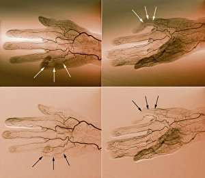
Background imageDiagnosis Collection: Blood supply of the hand, angiograms C018 / 0389
Blood supply of the hand, angiograms C018 / 0389
Blood supply of the hand. Coloured angiograms (X-rays) of the blood vessels of the right hand of a 65 year old patient with Dupuytrens contracture

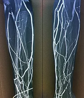
Background imageDiagnosis Collection: Forearm veins, X-ray C018 / 0475

Forearm veins, X-ray C018 / 0475
Forearm veins. Coloured venogram (vein X-ray) of the veins in the forearms of a 43 year old patient in need of dialysis. This venogram has been taken to evaluate the health of the veins prior to

Background imageDiagnosis Collection: Brain haemorrhage, CT scans C018 / 0569

Brain haemorrhage, CT scans C018 / 0569
Brain haemorrhage. Coloured computed tomography (CT) scans of the brain of a 49 year old patient with a subdural haematoma (blue)

Background imageDiagnosis Collection: Voiding cystourethrogram, X-rays C018 / 0577

Voiding cystourethrogram, X-rays C018 / 0577
Voiding cystourethrogram. X-ray of a 54 year old male patient undergoing a voiding cystourethrogram. This procedure involves liquid containing a radio-opaque dye being placed in the bladder via a

Background imageDiagnosis Collection: Healthy lumbar spine, MRI C018 / 0563

Healthy lumbar spine, MRI C018 / 0563
Healthy lumbar spine. Coloured magnetic resonance imaging (MRI) scan through the disc (red circle) between the second and third lumbar (lower back) vertebrae of a 30 year old patient

Background imageDiagnosis Collection: Healthy brain, MRI scan C018 / 0431

Healthy brain, MRI scan C018 / 0431
Healthy brain. Coloured 3D magnetic resonance imaging (MRI) scan of the healthy brain (excluding the brainstem) of a 37 year old. The cavities are the ventricles

Background imageDiagnosis Collection: Healthy brain, MRI scan C018 / 0430

Healthy brain, MRI scan C018 / 0430
Healthy brain. Coloured 3D magnetic resonance imaging (MRI) scan of the healthy brain (excluding the brainstem) of a 37 year old. The cavities are the ventricles

Background imageDiagnosis Collection: Uterine fibroids, MRI scan C018 / 0468

Uterine fibroids, MRI scan C018 / 0468
Uterine fibroids. Coloured magnetic resonance imaging (MRI) scan of the pelvis of a 47 year old female with two large fibroids (pink) in her uterus

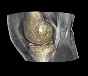
Background imageDiagnosis Collection: Knee injury, 3D CT scan C018 / 0648

Knee injury, 3D CT scan C018 / 0648
Knee injury. Coloured 3D computed tomography (CT) scan of a knee with a meniscal tear (white line at right between bones)

Background imageDiagnosis Collection: Voiding cystourethrogram, X-rays C018 / 0578

Voiding cystourethrogram, X-rays C018 / 0578
Voiding cystourethrogram. Coloured X-ray of a 54 year old male patient undergoing a voiding cystourethrogram. This procedure involves liquid containing a radio-opaque dye being placed in the bladder

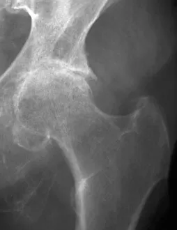
Background imageDiagnosis Collection: Osteoarthritis of the hip, X-ray C018 / 0593

Osteoarthritis of the hip, X-ray C018 / 0593
Osteoarthritis of the hip. X-ray the left hip of a 74 year old patient showing osteoarthritis. The ball at the top of the femur (thigh bone) fits into the socket of the hip

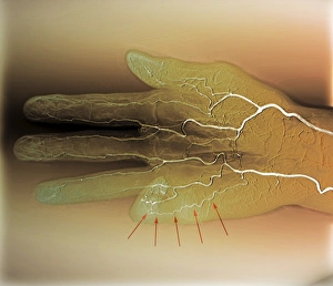
Background imageDiagnosis Collection: Blood supply of the hand, angiogram C018 / 0387

Blood supply of the hand, angiogram C018 / 0387
Blood supply of the hand. Coloured angiogram (X-ray) of the blood vessels of the right hand of a 65 year old patient with Dupuytrens contracture

Background imageDiagnosis Collection: Knee injury, 3D CT scan C018 / 0639

Knee injury, 3D CT scan C018 / 0639
Knee injury. Coloured 3D computed tomography (CT) scan of a knee with a meniscal tear (indicated by arrows). The menisci are fibrocartilage crescents that act as shock absorbers between the femur

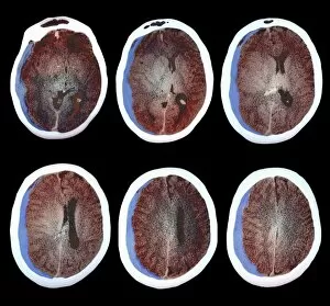
Background imageDiagnosis Collection: Hydrocephalus, CT scan C018 / 0568

Hydrocephalus, CT scan C018 / 0568
Hydrocephalus. Coloured computed tomography (CT) scan of the brain of a 22 year old patient with hydrocephalus (fluid in the brain)

Background imageDiagnosis Collection: Healthy brain, MRI scan C018 / 0429

Healthy brain, MRI scan C018 / 0429
Healthy brain. Coloured 3D magnetic resonance imaging (MRI) scan of the healthy brain (excluding the brainstem) of a 37 year old. The cavities are the ventricles

Background imageDiagnosis Collection: Fractured skull, X-ray C018 / 0591

Fractured skull, X-ray C018 / 0591
Fractured skull, X-ray. The fracture is the grey line at right that splits into two near the temple

Background imageDiagnosis Collection: Jeune syndrome, X-ray C018 / 0410

Jeune syndrome, X-ray C018 / 0410
Jeune syndrome. X-ray of the chest of a 2 year old with Jeune syndrome, also known as asphyxiating thoracic dystrophy. This inherited disease affects the development of the bones and cartilage

Background imageDiagnosis Collection: Cerebrotendinous xanthomatosis, MRI scan C018 / 0504

Cerebrotendinous xanthomatosis, MRI scan C018 / 0504
Cerebrotendinous xanthomatosis. Coloured magnetic resonance imaging (MRI) scan of the brain of a 46 year old patient with cerebrotendinous xanthomatosis (CTX)

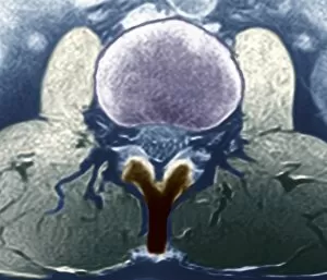
Background imageDiagnosis Collection: Healthy lumbar spine, MRI C018 / 0562

Healthy lumbar spine, MRI C018 / 0562
Healthy lumbar spine. Coloured magnetic resonance imaging (MRI) scan through the disc (purple) between the second and third lumbar (lower back) vertebrae of a 30 year old patient

Background imageDiagnosis Collection: Arthritic hands, X-ray C018 / 0534

Arthritic hands, X-ray C018 / 0534
Arthritic hands. Coloured X-ray of the hands of a 72 year old patient with rheumatoid arthritis. The wrist and finger joints have become inflamed and swollen

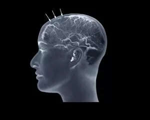
Background imageDiagnosis Collection: Thrombophlebitis, angiogram C018 / 0529

Thrombophlebitis, angiogram C018 / 0529
Thrombophlebitis. Coloured angiogram (X-ray) of the brain of a 28 year old female patient who had thrombophelibitis of the superior sagittal sinus (indicated by arrows)

Background imageDiagnosis Collection: Rectal cancer, MRI scans C018 / 0511

Rectal cancer, MRI scans C018 / 0511
Rectal cancer. Coloured magnetic resonance imaging (MRI) scans of the pelvis of a 65 year old patient with a malignant (cancerous) tumour (purple) in the rectum, the last part of the large intestine

Background imageDiagnosis Collection: Knee injury, 3D CT scan C018 / 0645

Knee injury, 3D CT scan C018 / 0645
Knee injury. Coloured 3D computed tomography (CT) scan of a knee with a meniscal tear (blue line at right between bones). The menisci are fibrocartilage crescents that act as shock absorbers between

Background imageDiagnosis Collection: Hydropneumothorax, X-ray C018 / 0586

Hydropneumothorax, X-ray C018 / 0586
Hydropneumothorax. Coloured X-ray of the chest of a 62 year old patient with a hydropneumothorax of the left lung (dark area at upper centre)

Background imageDiagnosis Collection: Colon cancer, barium X-ray C018 / 0574

Colon cancer, barium X-ray C018 / 0574
Colon cancer. Coloured X-ray of the large intestine of a 62 year old patient with cancer of the sigmoid colon. The patient was given a barium enema prior to the X-ray to show up any abnormalities

Background imageDiagnosis Collection: Uterine fibroids, MRI scan C018 / 0469

Uterine fibroids, MRI scan C018 / 0469
Uterine fibroids. Coloured magnetic resonance imaging (MRI) scan of the pelvis of a 47 year old female with two large fibroids (pink) in her uterus

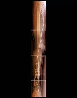
Background imageDiagnosis Collection: Pinned broken leg, angiogram C018 / 0392

Pinned broken leg, angiogram C018 / 0392
Pinned broken leg. Coloured angiogram of the right leg of a 22 year old patient that sustained an open tibial (shin bone) fracture in a motorcycle accident. The fracture has been aligned and pinned

Background imageDiagnosis Collection: Trachea, 3D CT scan C018 / 0393

Trachea, 3D CT scan C018 / 0393
Trachea. Coloured 3D computed tomography (CT) scan of the healthy trachea (windpipe) of a 45 year old patient at the point that it bifurcates into the two primary bronchi (airways)

Background imageDiagnosis Collection: Arthritic hands, X-ray C018 / 0532

Arthritic hands, X-ray C018 / 0532
Arthritic hands. X-ray of the hands of a 72 year old patient with rheumatoid arthritis. The wrist and finger joints have become inflamed and swollen

Background imageDiagnosis Collection: Hydrocephalus, CT scan C018 / 0567

Hydrocephalus, CT scan C018 / 0567
Hydrocephalus. Coloured computed tomography (CT) scan of the brain of a 22 year old patient with hydrocephalus (fluid in the brain)

Background imageDiagnosis Collection: Heart and lung disease, X-ray C018 / 0500

Heart and lung disease, X-ray C018 / 0500
Heart and lung disease. Coloured X-ray of the chest of an 80 year old patient with heart and lung disease. The patient is a former miner and has chronic obstructive pulmonary disease (COPD)

Background imageDiagnosis Collection: Hydrocephalus, CT scans C018 / 0566

Hydrocephalus, CT scans C018 / 0566
Hydrocephalus. Coloured computed tomography (CT) scans of the brain of a 22 year old patient with hydrocephalus (fluid in the brain)


All Professionally Made to Order for Quick Shipping

"Unlocking the Secrets: The Power of Diagnosis" A seahorse's hidden world revealed through an X-ray, reminding us that diagnosis goes beyond human health. Unveiling the spectrum: a color blindness test helping us understand how our eyes perceive the world differently. Eyesight test chart guiding us towards clarity and precision in diagnosing visual impairments. Peering into fractures: a broken wrist bone captured on X-ray, aiding doctors in accurate diagnosis and treatment plans. Exploring beneath the surface: a panoramic dental X-ray exposing hidden oral issues for comprehensive diagnoses. Empowering individuals with knowledge: home cholesterol test kits providing early detection for proactive health management. Saving lives through swift intervention: Pneumothorax treatment guided by an X-ray to restore lung function effectively. Knees tell stories too: a normal knee showcased on an X-ray, enabling precise diagnoses of joint conditions and injuries. Delving deeper into mysteries within: full body scans via MRI revealing intricate details for accurate medical assessments. Artistry meets science as ECGs portray the rhythm of a healthy heart, serving as diagnostic masterpieces in healthcare settings. Battling tuberculosis head-on with an X-ray, unveiling its presence so prompt treatments can be initiated promptly