Fraud Blocker Skip to main content

Diagnosis Collection (page 10)

"Unlocking the Secrets: The Power of Diagnosis" A seahorse's hidden world revealed through an X-ray, reminding us that diagnosis goes beyond human health

Background imageDiagnosis Collection: Hydropneumothorax, X-ray C018 / 0586

Hydropneumothorax, X-ray C018 / 0586
Hydropneumothorax. Coloured X-ray of the chest of a 62 year old patient with a hydropneumothorax of the left lung (dark area at upper centre)

Background imageDiagnosis Collection: Colon cancer, barium X-ray C018 / 0574

Colon cancer, barium X-ray C018 / 0574
Colon cancer. Coloured X-ray of the large intestine of a 62 year old patient with cancer of the sigmoid colon. The patient was given a barium enema prior to the X-ray to show up any abnormalities

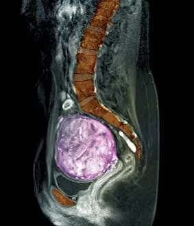
Background imageDiagnosis Collection: Uterine fibroids, MRI scan C018 / 0469

Uterine fibroids, MRI scan C018 / 0469
Uterine fibroids. Coloured magnetic resonance imaging (MRI) scan of the pelvis of a 47 year old female with two large fibroids (pink) in her uterus

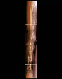
Background imageDiagnosis Collection: Pinned broken leg, angiogram C018 / 0392

Pinned broken leg, angiogram C018 / 0392
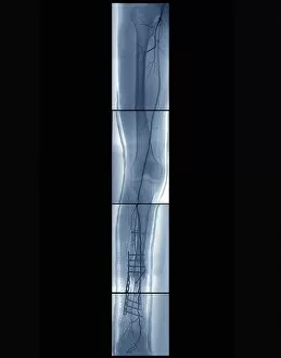
Pinned broken leg. Coloured angiogram of the right leg of a 22 year old patient that sustained an open tibial (shin bone) fracture in a motorcycle accident. The fracture has been aligned and pinned

Background imageDiagnosis Collection: Trachea, 3D CT scan C018 / 0393

Trachea, 3D CT scan C018 / 0393
Trachea. Coloured 3D computed tomography (CT) scan of the healthy trachea (windpipe) of a 45 year old patient at the point that it bifurcates into the two primary bronchi (airways)

Background imageDiagnosis Collection: Arthritic hands, X-ray C018 / 0532

Arthritic hands, X-ray C018 / 0532
Arthritic hands. X-ray of the hands of a 72 year old patient with rheumatoid arthritis. The wrist and finger joints have become inflamed and swollen

Background imageDiagnosis Collection: Hydrocephalus, CT scan C018 / 0567

Hydrocephalus, CT scan C018 / 0567
Hydrocephalus. Coloured computed tomography (CT) scan of the brain of a 22 year old patient with hydrocephalus (fluid in the brain)

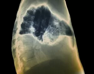
Background imageDiagnosis Collection: Heart and lung disease, X-ray C018 / 0500

Heart and lung disease, X-ray C018 / 0500
Heart and lung disease. Coloured X-ray of the chest of an 80 year old patient with heart and lung disease. The patient is a former miner and has chronic obstructive pulmonary disease (COPD)

Background imageDiagnosis Collection: Hydrocephalus, CT scans C018 / 0566

Hydrocephalus, CT scans C018 / 0566
Hydrocephalus. Coloured computed tomography (CT) scans of the brain of a 22 year old patient with hydrocephalus (fluid in the brain)

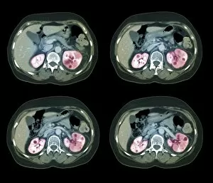
Background imageDiagnosis Collection: Kidney infection, CT scan C018 / 0473

Kidney infection, CT scan C018 / 0473
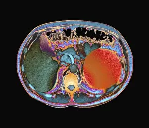
Kidney infection. Coloured computed tomography (CT) scans of the abdomen of a 57 year old patient with a kidney infection

Background imageDiagnosis Collection: Hydropneumothorax, X-ray C018 / 0585

Hydropneumothorax, X-ray C018 / 0585
Hydropneumothorax. Coloured X-ray of the chest of a 62 year old patient with a hydropneumothorax of the left lung (dark area at upper centre)

Background imageDiagnosis Collection: Early-onset Alzheimers disease, MRI scan

Early-onset Alzheimers disease, MRI scan
Early-onset Alzheimers disease. Magnetic resonance imaging (MRI) scans of the brain of a 53 year old patient with early-onset Alzheimers disease

Background imageDiagnosis Collection: Pinned broken leg, angiogram C018 / 0391

Pinned broken leg, angiogram C018 / 0391
Pinned broken leg. Coloured angiogram of the right leg of a 22 year old patient that sustained an open tibial (shin bone) fracture in a motorcycle accident. The fracture has been aligned and pinned

Background imageDiagnosis Collection: Rectal cancer, MRI scan C018 / 0512

Rectal cancer, MRI scan C018 / 0512
Rectal cancer. Coloured magnetic resonance imaging (MRI) scan of the pelvis of a 65 year old patient with a malignant (cancerous) tumour in the rectum, the last part of the large intestine

Background imageDiagnosis Collection: Benign dental tumour, CT scans C018 / 0589

Benign dental tumour, CT scans C018 / 0589
Benign dental tumour. Coloured computed tomography (CT) scans of the lower jaw of a 35 year old patient with a cemento-ossifying fibroma (purple)

Background imageDiagnosis Collection: Voiding cystourethrogram, X-rays C018 / 0579

Voiding cystourethrogram, X-rays C018 / 0579
Voiding cystourethrogram. Coloured X-ray of a 54 year old male patient undergoing a voiding cystourethrogram. This procedure involves liquid containing a radio-opaque dye being placed in the bladder

Background imageDiagnosis Collection: Jaw tumour, CT scan C018 / 0565

Jaw tumour, CT scan C018 / 0565
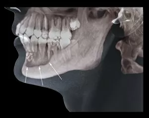
Jaw tumour. Coloured 3D computed tomography (CT) scan of the jaw of a 35 year old patient showing a benign (non-cancerous) bony lesion (indicated by arrows) in the mandible (lower jaw)

Background imageDiagnosis Collection: Healthy brain, MRI scan C018 / 0423

Healthy brain, MRI scan C018 / 0423
Healthy brain. Coloured 3D magnetic resonance imaging (MRI) scan of the healthy brain (excluding the brainstem) of a 37 year old. The cavities are the ventricles

Background imageDiagnosis Collection: Human head, MRI and 3D CT scans C018 / 0636

Human head, MRI and 3D CT scans C018 / 0636
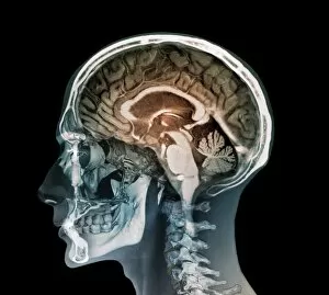
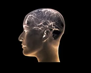
Human head. Coloured composite image of a magnetic resonance imaging (MRI) scan of the brain and 2D and 3D computed tomography (CT) scans of the head and neck of a 35 year old patient

Background imageDiagnosis Collection: Polycystic kidneys, CT scan C018 / 0573

Polycystic kidneys, CT scan C018 / 0573
Polycystic kidneys. Coloured computed tomography (CT) scan of the abdomen of a 28 year old patient with polycystic kidneys (dark blue)

Background imageDiagnosis Collection: Spinal cord cyst, MRI scan C018 / 0561

Spinal cord cyst, MRI scan C018 / 0561
Spinal cord cyst. Coloured magnetic resonance imaging (MRI) scan of the cervical (neck) spinal cord of a 28 year old patient with syringomyelia, a cyst in the spinal cord

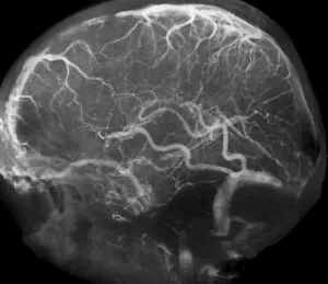
Background imageDiagnosis Collection: Thrombophlebitis, angiogram C018 / 0530

Thrombophlebitis, angiogram C018 / 0530
Thrombophlebitis. Coloured angiogram (X-ray) of the brain of a 28 year old female patient who had thrombophelibitis of the superior sagittal sinus (top left)

Background imageDiagnosis Collection: Voiding cystourethrogram, X-rays C018 / 0465

Voiding cystourethrogram, X-rays C018 / 0465
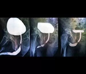
Voiding cystourethrogram. Three coloured X-rays showing a 65 year old patient undergoing a voiding cystourethrogram. This procedure involves liquid containing a radio-opaque dye being placed in

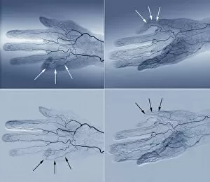
Background imageDiagnosis Collection: Blood supply of the hand, angiogram C018 / 0386
Blood supply of the hand, angiogram C018 / 0386
Blood supply of the hand. Coloured angiogram (X-ray) of the blood vessels of the right hand of a 65 year old patient with Dupuytrens contracture

Background imageDiagnosis Collection: Congenital kidney abnormality, X-ray C018 / 0516

Congenital kidney abnormality, X-ray C018 / 0516
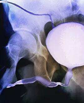
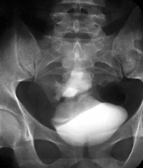
Congenital kidney abnormality. Coloured X-ray of the pelvis of a 28 year old patient who has only a single kidney (white area at lower centre) attached to the bladder (white area at bottom centre)

Background imageDiagnosis Collection: Blood supply of the hand, angiograms C018 / 0388

Blood supply of the hand, angiograms C018 / 0388
Blood supply of the hand. Coloured angiograms (X-rays) of the blood vessels of the right hand of a 65 year old patient with Dupuytrens contracture

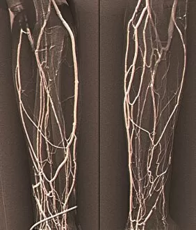
Background imageDiagnosis Collection: Forearm veins, X-ray C018 / 0474

Forearm veins, X-ray C018 / 0474
Forearm veins. Coloured venogram (vein X-ray) of the veins in the forearms of a 43 year old patient in need of dialysis. This venogram has been taken to evaluate the health of the veins prior to

Background imageDiagnosis Collection: Jeune syndrome, CT scans C018 / 0411

Jeune syndrome, CT scans C018 / 0411
Jeune syndrome. Coloured computed tomography (CT) scans of the chest of a 2 year old with Jeune syndrome, also known as asphyxiating thoracic dystrophy

Background imageDiagnosis Collection: Thrombophlebitis, angiogram C018 / 0528

Thrombophlebitis, angiogram C018 / 0528
Thrombophlebitis. Angiogram (X-ray) of the brain of a 28 year old female patient who had thrombophelibitis of the superior sagittal sinus (top left)

Background imageDiagnosis Collection: Bone growths, X-ray C018 / 0580

Bone growths, X-ray C018 / 0580
Bone growths. X-ray of the pelvis of a 12 year old patient with bony growths (exostoses) and cartilage cysts (enchondromas) on the femurs (thigh bones) and hip bones

Background imageDiagnosis Collection: Cardiology, conceptual image C017 / 7718

Cardiology, conceptual image C017 / 7718
MODEL RELEASED. Cardiology, conceptual image

Background imageDiagnosis Collection: Broken elbow, X-ray C017 / 7584

Broken elbow, X-ray C017 / 7584
Broken elbow. X-ray of the arm of a 55 year old male patient with fractures to the elbow and radius (right-most lower arm bone)

Background imageDiagnosis Collection: Knee surgery C016 / 9834

Knee surgery C016 / 9834
MODEL RELEASED. Knee surgery. Surgeon examining endoscopic images of a patients knee

Background imageDiagnosis Collection: Bladder stones, ultrasound scan C017 / 7261

Bladder stones, ultrasound scan C017 / 7261
Bladder stones. Ultrasound scan of the bladder of a 58 year old male showing bladder stones (bright white). Bladder stones (cystoliths)

Background imageDiagnosis Collection: Telemedicine, conceptual image C017 / 7597

Telemedicine, conceptual image C017 / 7597
Telemedicine, conceptual image

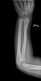
Background imageDiagnosis Collection: Broken arm, X-ray C017 / 7264

Broken arm, X-ray C017 / 7264
Broken arm. X-ray of the lower arm of a 6 year old male showing a greenstick fractures of the ulna (left) and radius (right) bones. Greenstick fractures occur when the bone bends and then cracks

Background imageDiagnosis Collection: Broken jaw, X-ray C017 / 7558

Broken jaw, X-ray C017 / 7558
Broken jaw. X-ray of the head of a 22 year old male patient with a fractured mandible (lower jaw bone). The fracture is seen at left

Background imageDiagnosis Collection: Arthritis of the neck, X-ray C017 / 7388

Arthritis of the neck, X-ray C017 / 7388
Arthritis of the neck. X-ray of a profile view of the neck of a 67 year old female with osteoarthritis of the neck. Osteoarthritis of the neck, also known as cervical spondylosis

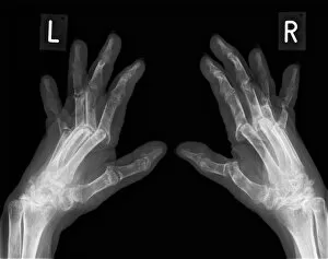
Background imageDiagnosis Collection: Arthritic hands, X-ray C017 / 7180

Arthritic hands, X-ray C017 / 7180
Arthritic hands, X-ray

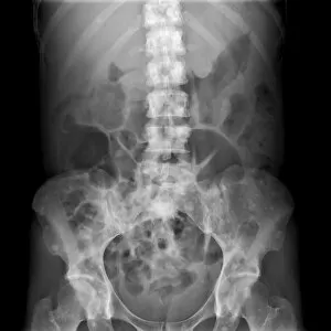
Background imageDiagnosis Collection: Secondary bone cancer, X-ray C017 / 7161

Secondary bone cancer, X-ray C017 / 7161
Secondary bone cancer. X-ray of the abdomen of a 40 year old female with metastatic (secondary) bone cancer (bright white areas). The cancer has metastasised (spread) from a primary cancer

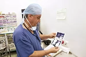
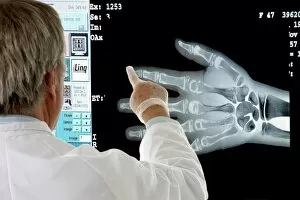
Background imageDiagnosis Collection: Radiologist examining a hand X-ray C014 / 1289

Radiologist examining a hand X-ray C014 / 1289
MODEL RELEASED. Radiologist examining a hand X-ray

Background imageDiagnosis Collection: Septic arthritis, X-ray C017 / 7385

Septic arthritis, X-ray C017 / 7385
Septic arthritis. X-ray of the shoulder joint of a 24 year old male that has been damaged due to septic arthritis. Septic arthritis is the inflammation of a joint due to a bacterial infection

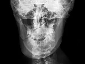
Background imageDiagnosis Collection: Facial fractures, X-ray C017 / 7638

Facial fractures, X-ray C017 / 7638
Facial fractures. X-ray of the skull of a 25 year old male patient with fractures to the right side (seen here on the left) of his face

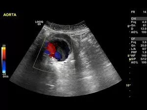
Background imageDiagnosis Collection: Abdominal aortic aneurysm, ultrasound C017 / 7646

Abdominal aortic aneurysm, ultrasound C017 / 7646
Abdominal aortic aneurysm. Doppler ultrasound scan of the abdomen of a 83 year old female patient with an abdominal aortic aneurysm (aA)

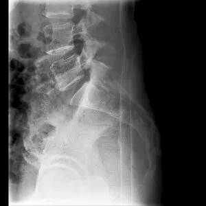
Background imageDiagnosis Collection: Fractured vertebra, X-ray C017 / 7585

Fractured vertebra, X-ray C017 / 7585
Fractured vertebra. X-ray of the lower spine of a 50 year old male with a fracture to the L1 lumbar vertebra (top centre)

Background imageDiagnosis Collection: Telemedicine, conceptual image C017 / 7414

Telemedicine, conceptual image C017 / 7414
Telemedicine, conceptual image

Background imageDiagnosis Collection: Telemedicine, conceptual image C017 / 7600

Telemedicine, conceptual image C017 / 7600
Telemedicine, conceptual image

Background imageDiagnosis Collection: Dislocated wrist, X-ray C017 / 7563

Dislocated wrist, X-ray C017 / 7563
Dislocated wrist. X-ray of the wrist of a 16 year old male that has been dislocated


All Professionally Made to Order for Quick Shipping

"Unlocking the Secrets: The Power of Diagnosis" A seahorse's hidden world revealed through an X-ray, reminding us that diagnosis goes beyond human health. Unveiling the spectrum: a color blindness test helping us understand how our eyes perceive the world differently. Eyesight test chart guiding us towards clarity and precision in diagnosing visual impairments. Peering into fractures: a broken wrist bone captured on X-ray, aiding doctors in accurate diagnosis and treatment plans. Exploring beneath the surface: a panoramic dental X-ray exposing hidden oral issues for comprehensive diagnoses. Empowering individuals with knowledge: home cholesterol test kits providing early detection for proactive health management. Saving lives through swift intervention: Pneumothorax treatment guided by an X-ray to restore lung function effectively. Knees tell stories too: a normal knee showcased on an X-ray, enabling precise diagnoses of joint conditions and injuries. Delving deeper into mysteries within: full body scans via MRI revealing intricate details for accurate medical assessments. Artistry meets science as ECGs portray the rhythm of a healthy heart, serving as diagnostic masterpieces in healthcare settings. Battling tuberculosis head-on with an X-ray, unveiling its presence so prompt treatments can be initiated promptly