Fraud Blocker Skip to main content

Diagnosis Collection (page 11)

"Unlocking the Secrets: The Power of Diagnosis" A seahorse's hidden world revealed through an X-ray, reminding us that diagnosis goes beyond human health

Background imageDiagnosis Collection: Facial fractures, X-ray C017 / 7638

Facial fractures, X-ray C017 / 7638
Facial fractures. X-ray of the skull of a 25 year old male patient with fractures to the right side (seen here on the left) of his face

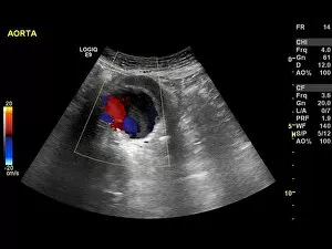
Background imageDiagnosis Collection: Abdominal aortic aneurysm, ultrasound C017 / 7646

Abdominal aortic aneurysm, ultrasound C017 / 7646
Abdominal aortic aneurysm. Doppler ultrasound scan of the abdomen of a 83 year old female patient with an abdominal aortic aneurysm (aA)

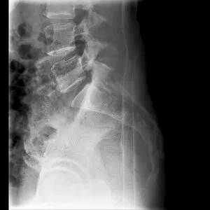
Background imageDiagnosis Collection: Fractured vertebra, X-ray C017 / 7585

Fractured vertebra, X-ray C017 / 7585
Fractured vertebra. X-ray of the lower spine of a 50 year old male with a fracture to the L1 lumbar vertebra (top centre)

Background imageDiagnosis Collection: Telemedicine, conceptual image C017 / 7414

Telemedicine, conceptual image C017 / 7414
Telemedicine, conceptual image

Background imageDiagnosis Collection: Telemedicine, conceptual image C017 / 7600

Telemedicine, conceptual image C017 / 7600
Telemedicine, conceptual image

Background imageDiagnosis Collection: Dislocated wrist, X-ray C017 / 7563

Dislocated wrist, X-ray C017 / 7563
Dislocated wrist. X-ray of the wrist of a 16 year old male that has been dislocated

Background imageDiagnosis Collection: Malunited fracture, X-ray C017 / 7641

Malunited fracture, X-ray C017 / 7641
Malunited fracture. X-ray of the leg of a 64 year old female patient with a malunited fracture of the femur (thigh bone). This occurs where a fracture fails to heal in the correct position

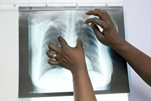
Background imageDiagnosis Collection: Tuberculosis X-ray diagnosis

Tuberculosis X-ray diagnosis
MODEL RELEASED. Tuberculosis (TB) X-ray diagnosis. Close-up of an X-ray of a patients lungs being examined by medical personnel for signs of TB

Background imageDiagnosis Collection: Urine diabetes test C014 / 1230

Urine diabetes test C014 / 1230
MODEL RELEASED. Urine diabetes test. Woman using a urine self-test for diabetes. This test detects the levels of glucose and ketone in the urine

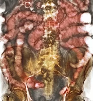
Background imageDiagnosis Collection: Small bowel obstruction, X-ray C014 / 1819

Small bowel obstruction, X-ray C014 / 1819
Small bowel obstruction. Coloured X-ray of the abdomen of a 77 year old male patient with a small bowel obstruction and gallstones (spheres at top left). The white areas are gas-filled bowel loops

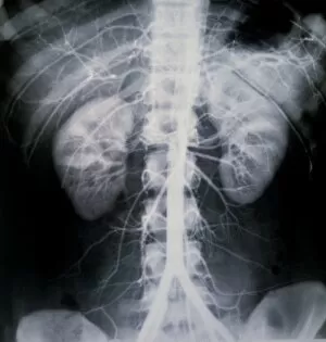
Background imageDiagnosis Collection: Healthy kidneys, X-ray C014 / 4920

Healthy kidneys, X-ray C014 / 4920
Healthy kidneys. Arteriograph (artery X-ray) of the abdomen showing two healthy kidneys (yellow, left and right). he kidneys filter waste products from the blood

Background imageDiagnosis Collection: Uterine fibroid, X-ray C014 / 4919

Uterine fibroid, X-ray C014 / 4919
Uterus tumour. Coloured X-ray of the abdomen of a woman with a fibroid (round, centre) in her uterus. A fibroid is a fibrous benign tumour originating from muscular tissue (leimyoma)

Background imageDiagnosis Collection: Uterine fibroid, X-ray C014 / 4918

Uterine fibroid, X-ray C014 / 4918
Uterus tumour. Coloured X-ray of the abdomen of a woman with a fibroid (orange, round) in her uterus. A fibroid is a fibrous benign tumour originating from muscular tissue (leimyoma)

Background imageDiagnosis Collection: Endometriosis, X-ray C014 / 4917

Endometriosis, X-ray C014 / 4917
Endometriosis. Coloured X-ray of the abdomen of woman with endometriosis. Endometriosis is the movement of cells from the lining of the uterus to the abdominal cavity

Background imageDiagnosis Collection: Patient undergoing doppler ultrasound

Patient undergoing doppler ultrasound
Doppler ultrasound. A patient undergoing a doppler ultrasound investigation of the common carotid artery in the neck. Doppler ultrasound gives a view of the speed of blood flow in a vessel

Background imageDiagnosis Collection: Endometriosis, X-ray C014 / 4916

Endometriosis, X-ray C014 / 4916
Endometriosis. Coloured X-ray of the abdomen of woman with endometriosis. Endometriosis is the movement of cells from the lining of the uterus to the abdominal cavity

Background imageDiagnosis Collection: Healthy kidneys, X-ray C014 / 4921

Healthy kidneys, X-ray C014 / 4921
Healthy kidneys. Arteriograph (artery X-ray) of the abdomen showing two healthy kidneys (orange). he kidneys filter waste products from the blood

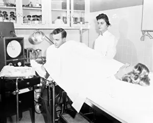
Background imageDiagnosis Collection: Rubin gynaecology test, 1956

Rubin gynaecology test, 1956
Rubin gynaecology test. Doctor with nurse and patient in a hospital, with the doctor carrying out a test on the female patients fallopian tubes

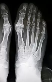
Background imageDiagnosis Collection: Hallux rigidus before surgery, X-ray

Hallux rigidus before surgery, X-ray
Hallux rigidus before surgery. X-ray showing foot bones before surgery to correct a case of hallux rigidus, a stiff big toe

Background imageDiagnosis Collection: Colonic spasm, X-ray

Colonic spasm, X-ray
Colonic spasm. Double contrast coloured X-ray of a spasm in the large intestine (colon). The spasm is the narrowed area at left. This could be caused by irritable bowel syndrome

Background imageDiagnosis Collection: Stomach cancer, barium X-ray

Stomach cancer, barium X-ray
Stomach cancer. Coloured frontal X-ray of the upper gastroduodenal tract of a patient with stomach cancer (grey, upper centre)

Background imageDiagnosis Collection: Atherosclerosis, X-ray

Atherosclerosis, X-ray
Atherosclerosis. X-ray of a patient with atherosclerosis of the abdominal aorta (white, down centre). The aorta is the bodys main artery, it carries blood from the heart to the rest of the body

Background imageDiagnosis Collection: Bowel disease in the colon C016 / 8338

Bowel disease in the colon C016 / 8338
Bowel disease in the colon. Endoscopic view of diverticular disease (diverticulosis) affecting the sigmoid colon. Diverticulosis causes sacs (diverticula)

Background imageDiagnosis Collection: Secondary bowel cancer, endoscopic view C016 / 8337

Secondary bowel cancer, endoscopic view C016 / 8337
Secondary bowel cancer. Endoscopic view of the interior of a patients descending duodenum (first part of the small intestine), showing cancer that has spread (metastasised) from the pancreas

Background imageDiagnosis Collection: Intestinal lymphoid hyperplasia C016 / 8334

Intestinal lymphoid hyperplasia C016 / 8334
Intestinal lymphoid hyperplasia. Endoscopic view of lymphoid hyperplasia in the terminal ileum - the last part of the small intestine

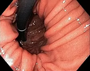
Background imageDiagnosis Collection: Hiatal hernia, endoscopic view C016 / 8330

Hiatal hernia, endoscopic view C016 / 8330
Hiatal hernia. Endoscopic view of a hiatal hernia (gastro-oesophageal prolapse), seen from below, with the endoscope(black) seen in an inverted position inside the stomach

Background imageDiagnosis Collection: Hiatal hernia, endoscopic view C016 / 8329

Hiatal hernia, endoscopic view C016 / 8329
Hiatal hernia. Endoscopic view of a hiatal hernia (gastro-oesophageal prolapse), seen from above. A hernia is the protrusion of an organ, or the fascia of an organ

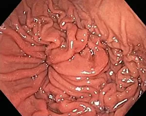
Background imageDiagnosis Collection: Hyperplastic polyps in the stomach C016 / 8327

Hyperplastic polyps in the stomach C016 / 8327
Hyperplastic polyps in the stomach. Endoscopic view of small hyperplastic polyps (growths) in the gastric fundus of the stomach

Background imageDiagnosis Collection: Diverticulum of the duodenum C016 / 8320

Diverticulum of the duodenum C016 / 8320
Diverticulum of the duodenum. Endoscope view of a diverticulum in the wall of the descending part of the duodenum - the first section of the small intestine

Background imageDiagnosis Collection: Crohns disease, endoscopic view

Crohns disease, endoscopic view
Crohns disease. Endoscopic view of the interior of a patients neoterminal ileum affected by Crohns disease, showing ulceration (white)

Background imageDiagnosis Collection: Ulcerative colitis, endoscopic view C016 / 8313

Ulcerative colitis, endoscopic view C016 / 8313
Ulcerative colitis. Endoscopic view of the interior of a patients distal colon (large intestine) affected by ulcerative colitis (UC), showing ulceration (white areas)

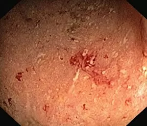
Background imageDiagnosis Collection: Antrum occult mucosal bleeding C016 / 8314

Antrum occult mucosal bleeding C016 / 8314
Antrum occult mucosal bleeding. Endoscopic view of the interior of a patients antrum (final portion of stomach), showing bleeding (red) of the mucosal lining

Background imageDiagnosis Collection: Ulcerative colitis, endoscopic view C016 / 8312

Ulcerative colitis, endoscopic view C016 / 8312
Ulcerative colitis. Endoscopic view of the interior of a patients distal colon (large intestine) affected by ulcerative colitis (UC), showing ulceration (white areas)

Background imageDiagnosis Collection: Ulcerative proctitis, endoscopic view C016 / 8311

Ulcerative proctitis, endoscopic view C016 / 8311
Ulcerative colitis. Endoscopic view of the interior of a patients anus, showing ulceration (white areas) cause by ulcerative proctitis

Background imageDiagnosis Collection: Ulcerative colitis, endoscopic view C016 / 8310

Ulcerative colitis, endoscopic view C016 / 8310
Ulcerative colitis. Endoscopic view of the interior of a patients colon (large intestine) affected by ulcerative colitis (UC), showing ulceration (white areas)

Background imageDiagnosis Collection: Ulcerative colitis, endoscopic view C016 / 8309

Ulcerative colitis, endoscopic view C016 / 8309
Ulcerative colitis. Endoscopic view of the interior of a patients colon (large intestine) affected by ulcerative colitis (UC), showing ulceration (white areas)

Background imageDiagnosis Collection: Ulcerative colitis, endoscopic view C016 / 8306

Ulcerative colitis, endoscopic view C016 / 8306
Ulcerative colitis. Endoscopic view of the interior of a patients distal colon (large intestine) affected by ulcerative colitis (UC), showing ulceration (white areas)

Background imageDiagnosis Collection: Colon cancer, endoscopic view C016 / 8307

Colon cancer, endoscopic view C016 / 8307
Colon cancer. Endoscopic view of a malignant (cancerous) adenocarcinoma (lower right) in a patients ascending colon (large intestine)

Background imageDiagnosis Collection: Melanosis coli, endoscopic view C016 / 8304

Melanosis coli, endoscopic view C016 / 8304
Melanosis coli. Endoscopic view of the interior of a patients colon affected by melanosis coli (pseudomelanosis coli). This benign condition causes brown pigmentation of the lining of the colon

Background imageDiagnosis Collection: Brain injury, MRI scan

Brain injury, MRI scan
Brain injury. Coronal magnetic resonance imaging (MRI) brain scan showing intracranial abnormalities in a 23-year-old male motorcycle crash victim

Background imageDiagnosis Collection: Digital thermometer showing fever C014 / 0109

Digital thermometer showing fever C014 / 0109
MODEL RELEASED. Digital thermometer showing fever, held in a teenage girls hand. The temperature reading of 38.60 degrees Celsius is elevated above normal human body temperature

Background imageDiagnosis Collection: Eyesight test, conceptual image

Eyesight test, conceptual image. Composite image of a human eye and an eyesight test chart

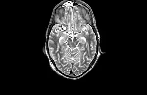
Background imageDiagnosis Collection: MRI scan

MRI scan

Background imageDiagnosis Collection: Endometrial hyperplasia, light micrograph C015 / 7104

Endometrial hyperplasia, light micrograph C015 / 7104
Endometrial hyperplasia. Light micrograph of a section through tissue from the endometrium, in a case of endometrial hyperplasia. The endometrium is the uterus lining

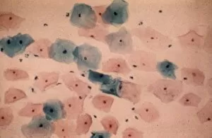
Background imageDiagnosis Collection: Healthy cervical smear, micrograph C015 / 6995

Healthy cervical smear, micrograph C015 / 6995
Healthy cervical smear. Light micrograph of healthy squamous epithelial cells from a smear test. A cervical smear, or pap test (after the papanicolaou used to stain the slides)

Background imageDiagnosis Collection: Cancer of the chorion C015 / 6905

Cancer of the chorion C015 / 6905
Cancer of the chorion. Light micrograph of a section through a sarcoma of the chorion, a malignant (cancerous) tumour of the membrane (chorion) that surrounds a developing embryo

Background imageDiagnosis Collection: Cervical smear showing endometrial cancer C015 / 6750

Cervical smear showing endometrial cancer C015 / 6750
Cervical smear showing endometrial cancer. Light micrograph of cells from a cervical smear test showing endometrial sarcoma. This is a cancer of the lining of the womb, the endometrium

Background imageDiagnosis Collection: Fractured and dislocated shoulder, X-ray C016 / 6607

Fractured and dislocated shoulder, X-ray C016 / 6607
Fractured and dislocated shoulder. Coloured X-ray of the shoulder of a 67 year old patient who has fallen and fractured the neck of the humerus (upper arm bone) and dislocated the shoulder


All Professionally Made to Order for Quick Shipping

"Unlocking the Secrets: The Power of Diagnosis" A seahorse's hidden world revealed through an X-ray, reminding us that diagnosis goes beyond human health. Unveiling the spectrum: a color blindness test helping us understand how our eyes perceive the world differently. Eyesight test chart guiding us towards clarity and precision in diagnosing visual impairments. Peering into fractures: a broken wrist bone captured on X-ray, aiding doctors in accurate diagnosis and treatment plans. Exploring beneath the surface: a panoramic dental X-ray exposing hidden oral issues for comprehensive diagnoses. Empowering individuals with knowledge: home cholesterol test kits providing early detection for proactive health management. Saving lives through swift intervention: Pneumothorax treatment guided by an X-ray to restore lung function effectively. Knees tell stories too: a normal knee showcased on an X-ray, enabling precise diagnoses of joint conditions and injuries. Delving deeper into mysteries within: full body scans via MRI revealing intricate details for accurate medical assessments. Artistry meets science as ECGs portray the rhythm of a healthy heart, serving as diagnostic masterpieces in healthcare settings. Battling tuberculosis head-on with an X-ray, unveiling its presence so prompt treatments can be initiated promptly