Fraud Blocker Skip to main content

Diagnosis Collection (page 8)

"Unlocking the Secrets: The Power of Diagnosis" A seahorse's hidden world revealed through an X-ray, reminding us that diagnosis goes beyond human health

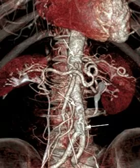
Background imageDiagnosis Collection: Atherosclerosis, 3D CT scan C016 / 6540

Atherosclerosis, 3D CT scan C016 / 6540
Atherosclerosis. Coloured 3D computed tomography (CT) angiogram of the abdomen of a 73 year old patient. The aorta (cream, down centre), the bodys main artery, has calcified fatty plaques (atheroma)

Background imageDiagnosis Collection: Light microscopy C016 / 9700

Light microscopy C016 / 9700
Light microscopy. Light microscope being used to magnify a blood sample

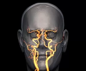
Background imageDiagnosis Collection: Atherosclerosis, 3D CT scan C016 / 6499

Atherosclerosis, 3D CT scan C016 / 6499
Atherosclerosis. Coloured 3D computed tomography (CT) angiogram of the head of a 60 year old patient with hypertension (high blood pressure) and hypercholesterolemia (high blood cholesterol)

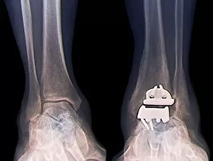
Background imageDiagnosis Collection: Replacement ankle joint, X-ray C016 / 6583

Replacement ankle joint, X-ray C016 / 6583
Replacement ankle joint. Coloured X-ray of the ankles of a 59 year old patient showing a prosthetic ankle joint between the left tibia (shin bone) and talus (ankle bone)

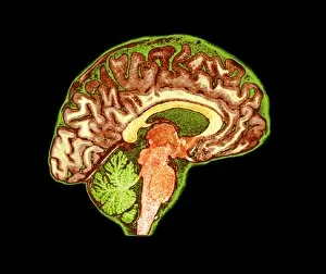
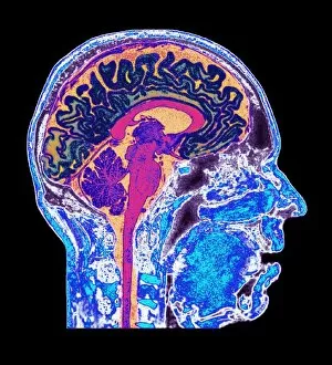
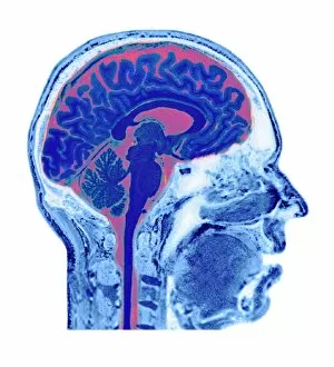
Background imageDiagnosis Collection: Normal human brain, MRI scan C016 / 8837

Normal human brain, MRI scan C016 / 8837
Brain. Coloured magnetic resonance imaging (MRI) scan of a sagittal section through a healthy human brain. The highly folded area (orange) is the cerebrum, which is responsible for conscious thought
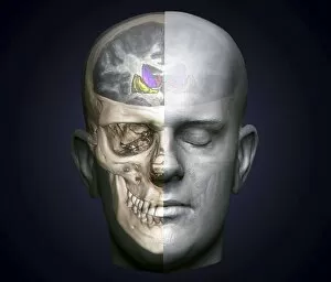
Background imageDiagnosis Collection: Human head, MRI and 3D CT scans C016 / 6409

Human head, MRI and 3D CT scans C016 / 6409
Human head. Coloured composite image of a magnetic resonance imaging (MRI) scan of the brain and a 3D computed tomography (CT) scan of the skull of a 35 year old patient

Background imageDiagnosis Collection: Human head, MRI and 3D CT scans C016 / 6416

Human head, MRI and 3D CT scans C016 / 6416
Human head. Coloured composite image of a magnetic resonance imaging (MRI) scan of the brain and 2D and 3D computed tomography (CT) scans of the head and shoulders of a 38 year old patient

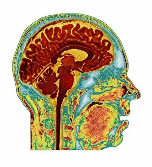
Background imageDiagnosis Collection: Normal human brain, MRI scan C016 / 8841

Normal human brain, MRI scan C016 / 8841
Brain. Coloured magnetic resonance imaging (MRI) scan of a sagittal section through a healthy human brain. The highly folded area (red and yellow) is the cerebrum

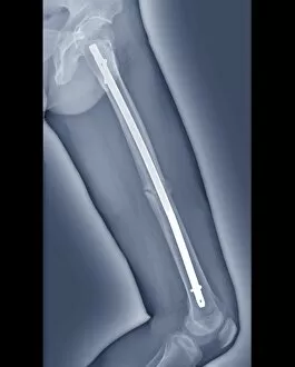
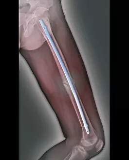
Background imageDiagnosis Collection: Pinned thigh fracture, X-ray C016 / 6563

Pinned thigh fracture, X-ray C016 / 6563
Pinned thigh fracture. Coloured X-ray of the fractured femur (thigh bone) of a 52 year old patient. The broken bone has been fixed internally with a metal rod

Background imageDiagnosis Collection: Healthy lungs, 3D CT scan C016 / 6497

Healthy lungs, 3D CT scan C016 / 6497
Healthy lungs. Coloured 3D computed tomography (CT) scan of the lungs of a 37 year old patient

Background imageDiagnosis Collection: Healthy lungs, 3D CT scan C016 / 6503

Healthy lungs, 3D CT scan C016 / 6503
Healthy lungs. Coloured 3D computed tomography (CT) scan of the chest of a 54 year old patient showing healthy lungs. At centre is the aortic arch (red), where the aorta, the bodys main artery

Background imageDiagnosis Collection: Light microscopy C016 / 9703

Light microscopy C016 / 9703
Light microscopy. Light microscope being used to magnify a blood sample

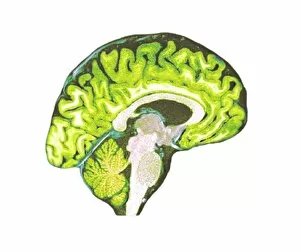
Background imageDiagnosis Collection: Normal human brain, MRI scan C016 / 8835

Normal human brain, MRI scan C016 / 8835
Brain. Coloured magnetic resonance imaging (MRI) scan of a sagittal section through a healthy human brain. The highly folded area across top is the cerebrum

Background imageDiagnosis Collection: Healthy lungs, 3D CT scan C016 / 6502

Healthy lungs, 3D CT scan C016 / 6502
Healthy lungs. Coloured 3D computed tomography (CT) scan of the chest of a 54 year old patient showing healthy lungs

Background imageDiagnosis Collection: Normal human brain, MRI scan C016 / 8846

Normal human brain, MRI scan C016 / 8846
Brain. Coloured magnetic resonance imaging (MRI) scan of a sagittal section through a patients head showing a healthy human brain

Background imageDiagnosis Collection: Healthy lungs, 3D CT scan C016 / 6506

Healthy lungs, 3D CT scan C016 / 6506
Healthy lungs. Coloured 3D computed tomography (CT) scan of the chest of a 35 year old patient showing healthy lungs

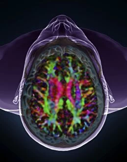
Background imageDiagnosis Collection: Human head and brain, DTI and 3D CT scans C016 / 6411

Human head and brain, DTI and 3D CT scans C016 / 6411
Brain cancer. Coloured composite image of a diffusion tensor imaging (DTI) scan of the brain and a 3D computed tomography (CT)

Background imageDiagnosis Collection: Normal human brain, MRI scan C016 / 8834

Normal human brain, MRI scan C016 / 8834
Brain. Coloured magnetic resonance imaging (MRI) scan of a sagittal section through a healthy human brain. The highly folded area (orange) is the cerebrum, which is responsible for conscious thought

Background imageDiagnosis Collection: Healthy lungs, 3D CT scan C016 / 6495

Healthy lungs, 3D CT scan C016 / 6495
Healthy lungs. Coloured 3D computed tomography (CT) scan of the chest of a 37 year old patient showing healthy lungs

Background imageDiagnosis Collection: Normal human brain, MRI scan C016 / 8849

Normal human brain, MRI scan C016 / 8849
Brain. Coloured magnetic resonance imaging (MRI) scan of a sagittal section through a patients head showing a healthy human brain

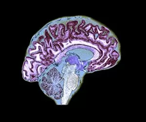
Background imageDiagnosis Collection: Normal human brain, MRI scan C016 / 8836

Normal human brain, MRI scan C016 / 8836
Brain. Coloured magnetic resonance imaging (MRI) scan of a sagittal section through a healthy human brain. The highly folded area across top is the cerebrum

Background imageDiagnosis Collection: Atherosclerosis, 3D CT scan C016 / 6538

Atherosclerosis, 3D CT scan C016 / 6538
Atherosclerosis. Coloured 3D computed tomography (CT) angiogram of the hip of a 67 year old patient with hypertension (high blood pressure) and hypercholesterolemia (high blood cholesterol)

Background imageDiagnosis Collection: Normal human brain, MRI scan C016 / 8844

Normal human brain, MRI scan C016 / 8844
Brain. Coloured magnetic resonance imaging (MRI) scan of a sagittal section through a patients head showing a healthy human brain

Background imageDiagnosis Collection: Atherosclerosis, 3D CT scan C016 / 6539

Atherosclerosis, 3D CT scan C016 / 6539
Atherosclerosis. Coloured 3D computed tomography (CT) angiogram of the heart of a 69 year old patient that has had an aortic heart valve replacement

Background imageDiagnosis Collection: Healthy lungs, 3D CT scan C016 / 6498

Healthy lungs, 3D CT scan C016 / 6498
Healthy lungs. Coloured 3D computed tomography (CT) scan of the lungs of a 37 year old patient

Background imageDiagnosis Collection: Human head, MRI and 3D CT scans C016 / 6418

Human head, MRI and 3D CT scans C016 / 6418
Human head. Coloured composite image of a magnetic resonance imaging (MRI) scan of the brain and 2D and 3D computed tomography (CT) scans of the head and shoulders of a 38 year old patient

Background imageDiagnosis Collection: Human head and brain, DTI and 3D CT scans C016 / 6410

Human head and brain, DTI and 3D CT scans C016 / 6410
Human head. Coloured composite image of a diffusion tensor imaging (DTI) scan of the brain and a 3D computed tomography (CT) scan of the skull of a 35 year old patient

Background imageDiagnosis Collection: Normal human brain, MRI scan C016 / 8839

Normal human brain, MRI scan C016 / 8839
Brain. Coloured magnetic resonance imaging (MRI) scan of a sagittal section through a healthy human brain. The highly folded area across top is the cerebrum

Background imageDiagnosis Collection: Pinned thigh fracture, X-ray C016 / 6564

Pinned thigh fracture, X-ray C016 / 6564
Pinned thigh fracture. Coloured X-ray of the fractured femur (thigh bone) of a 52 year old patient. The broken bone has been fixed internally with a metal rod

Background imageDiagnosis Collection: Scoliosis of the spine, X-ray C016 / 6565

Scoliosis of the spine, X-ray C016 / 6565
Scoliosis of the spine. Coloured X-ray of the chest of a 34 year old female patient with scoliosis (sideways curvature) of the spine

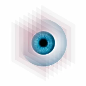
Background imageDiagnosis Collection: Diagnostics imaging techniques, concept C016 / 7511

Diagnostics imaging techniques, concept C016 / 7511
Diagnostics imaging techniques, conceptual computer artwork

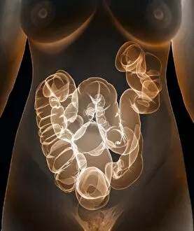
Background imageDiagnosis Collection: Healthy large intestine, 3D CT scan C016 / 6508

Healthy large intestine, 3D CT scan C016 / 6508
Healthy large intestine. Coloured 3D computed tomography (CT) scan of the abdomen of a 37 year old female patient showing a healthy large intestine

Background imageDiagnosis Collection: Healthy large intestine, 3D CT scan C016 / 6507

Healthy large intestine, 3D CT scan C016 / 6507
Healthy large intestine. Coloured 3D computed tomography (CT) scan of the abdomen of a 37 year old female patient showing a healthy large intestine

Background imageDiagnosis Collection: Human head, MRI and 3D CT scans C016 / 6396

Human head, MRI and 3D CT scans C016 / 6396
Human head. Coloured composite image of a magnetic resonance imaging (MRI) scan of the brain and a 3D computed tomography (CT) scan of the skull of a 35 year old patient

Background imageDiagnosis Collection: Light microscopy C016 / 9702

Light microscopy C016 / 9702
Light microscopy. Light microscope being used to magnify a blood sample

Background imageDiagnosis Collection: Human head, MRI and 3D CT scans C016 / 6398

Human head, MRI and 3D CT scans C016 / 6398
Human head. Coloured composite image of a magnetic resonance imaging (MRI) scan of the brain and a 3D computed tomography (CT) scan of the skull of a 35 year old patient

Background imageDiagnosis Collection: Normal human brain, MRI scan C016 / 8847

Normal human brain, MRI scan C016 / 8847
Brain. Coloured magnetic resonance imaging (MRI) scan of a sagittal section through a patients head showing a healthy human brain

Background imageDiagnosis Collection: Human head, MRI and 3D CT scans C016 / 6395

Human head, MRI and 3D CT scans C016 / 6395
Human head. Coloured composite image of a magnetic resonance imaging (MRI) scan of the brain and a 3D computed tomography (CT) scan of the skull of a 35 year old patient

Background imageDiagnosis Collection: Healthy lungs, 3D CT scan C016 / 6505

Healthy lungs, 3D CT scan C016 / 6505
Healthy lungs. Coloured 3D computed tomography (CT) scan of the chest of a 35 year old patient showing healthy lungs

Background imageDiagnosis Collection: Human head, MRI and 3D CT scans C016 / 6494

Human head, MRI and 3D CT scans C016 / 6494
Human head. Coloured composite image of a magnetic resonance imaging (MRI) scan of the brain and a 3D computed tomography (CT) scan of the face of a 40 year old patient

Background imageDiagnosis Collection: Light microscopy C016 / 9701

Light microscopy C016 / 9701
Light microscopy. Light microscope being used to magnify a blood sample

Background imageDiagnosis Collection: Human head, MRI and 3D CT scans C016 / 6415

Human head, MRI and 3D CT scans C016 / 6415
Stroke. Coloured composite image of a magnetic resonance imaging (MRI) scan of the brain and 2D and 3D computed tomography (CT)

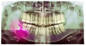
Background imageDiagnosis Collection: Impacted wisdom tooth, panoral X-ray

Impacted wisdom tooth, panoral X-ray
Impacted wisdom tooth. Coloured panoral X-ray of the mouth of a patient with an impacted wisdom tooth (pink) in the right lower jaw

Background imageDiagnosis Collection: Womb, CT scan

Womb, CT scan
Coloured computed tomography (CT) scan in sagittal (side) section through a womans lower abdomen showing the uterus (womb, pink)

Background imageDiagnosis Collection: Normal skull, 3D CT scan C016 / 6330

Normal skull, 3D CT scan C016 / 6330
Normal skull. Coloured cut-away 3D computed tomography (CT) scan of the healthy skull of a 35 year old patient

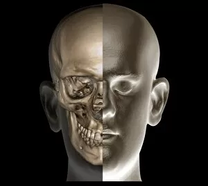
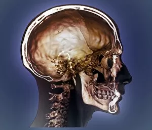
Background imageDiagnosis Collection: Human head, 3D CT scan C016 / 6339

Human head, 3D CT scan C016 / 6339
Human head. Coloured 3D computed tomography (CT) scan of a healthy human head. The soft tissues are shown at right, the skull is seen at left

Background imageDiagnosis Collection: Normal head and neck, 3D CT scan C016 / 6335

Normal head and neck, 3D CT scan C016 / 6335
Normal head. Coloured 3D computed tomography (CT) scan of the healthy head and neck of a 35 year old patient

Background imageDiagnosis Collection: Normal head and neck, 3D CT scan C016 / 6334

Normal head and neck, 3D CT scan C016 / 6334
Normal head. Coloured 3D computed tomography (CT) scan of the healthy head and neck of a 35 year old patient


All Professionally Made to Order for Quick Shipping

"Unlocking the Secrets: The Power of Diagnosis" A seahorse's hidden world revealed through an X-ray, reminding us that diagnosis goes beyond human health. Unveiling the spectrum: a color blindness test helping us understand how our eyes perceive the world differently. Eyesight test chart guiding us towards clarity and precision in diagnosing visual impairments. Peering into fractures: a broken wrist bone captured on X-ray, aiding doctors in accurate diagnosis and treatment plans. Exploring beneath the surface: a panoramic dental X-ray exposing hidden oral issues for comprehensive diagnoses. Empowering individuals with knowledge: home cholesterol test kits providing early detection for proactive health management. Saving lives through swift intervention: Pneumothorax treatment guided by an X-ray to restore lung function effectively. Knees tell stories too: a normal knee showcased on an X-ray, enabling precise diagnoses of joint conditions and injuries. Delving deeper into mysteries within: full body scans via MRI revealing intricate details for accurate medical assessments. Artistry meets science as ECGs portray the rhythm of a healthy heart, serving as diagnostic masterpieces in healthcare settings. Battling tuberculosis head-on with an X-ray, unveiling its presence so prompt treatments can be initiated promptly