Canvas Print : Muscle anatomy


![]()
Canvas Prints from Science Photo Library
Muscle anatomy
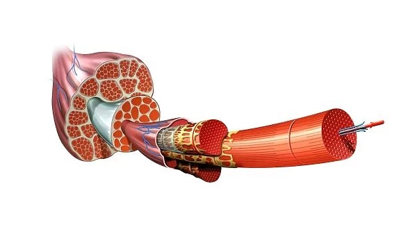
Muscle anatomy. Cutaway artwork showing the anatomy and internal structure of a muscle, from the macroscopic (left) to the microscopic (right) level. At upper left, a muscle is shown with nerves (blue). The cutaway section shows the muscle consisting of muscle bundles (fascicles). Each fascicle consists of muscle fibres (the third stage shown here, also with nerves, blue). The fibres consist of myofibrils, shown here with green and yellow sheaths, the cell membrane or sarcolemma. The smallest element are the myofilaments (right) consisting of the proteins actin (blue) and myosin (red)
Science Photo Library features Science and Medical images including photos and illustrations
Media ID 6317425
© JOSE ANTONIO PEAS/SCIENCE PHOTO LIBRARY
Actin Bundle Bundles Cut Away Exploded Fascicle Fiber Fibre Fibres Filament Filaments Magnified Membrane Microscopic Muscles Myofibril Myofibrils Myofilament Myology Myosin Nerve Nerves Proteins Scale Macroscopic Protein Section Sectioned
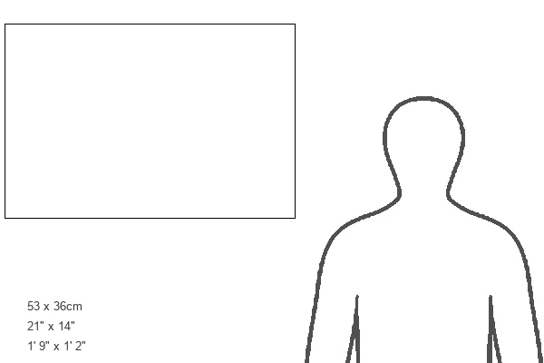
21"x14" (53x35cm) Canvas Print
Explore the intricacies of muscle anatomy with our stunning Canvas Prints from Media Storehouse, featuring this captivating image from Science Photo Library. Witness the transformation from the macroscopic to the microscopic level, as this cutaway artwork reveals the complex internal structure of a muscle. Bring the wonders of science into your home or office with our high-quality, vibrant Canvas Prints. Perfect for educational settings or for those with a deep appreciation for the human body, this Muscle Anatomy print is sure to impress.
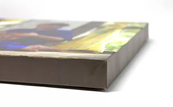
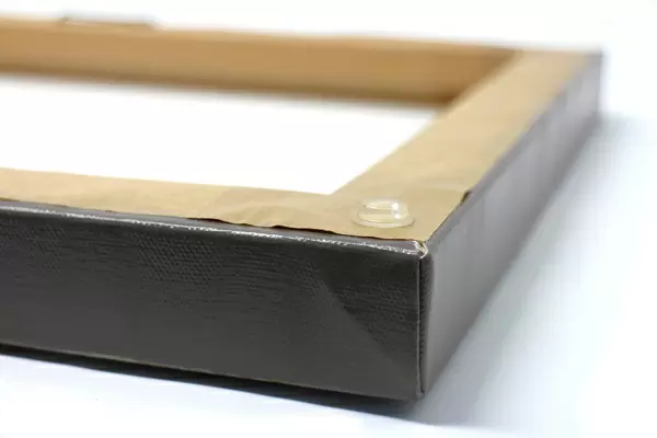
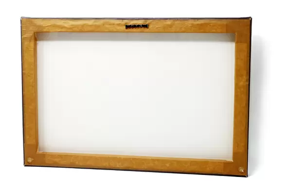
Ready to hang Premium Gloss Canvas Print. Our archival quality canvas prints are made from Polyester and Cotton mix and stretched over a 1.25" (32mm) kiln dried knot free wood stretcher bar. Packaged in a plastic bag and secured to a cardboard insert for transit.
Canvas Prints add colour, depth and texture to any space. Professionally Stretched Canvas over a hidden Wooden Box Frame and Ready to Hang
Estimated Image Size (if not cropped) is 53.3cm x 29.1cm (21" x 11.5")
Estimated Product Size is 53.3cm x 35.6cm (21" x 14")
These are individually made so all sizes are approximate
Artwork printed orientated as per the preview above, with landscape (horizontal) orientation to match the source image.
EDITORS COMMENTS
This print from Science Photo Library showcases the intricate and complex anatomy of a muscle. The artwork provides a cutaway view, revealing the internal structure of the muscle at both macroscopic and microscopic levels. On the left side of the image, we see a muscle with its accompanying nerves depicted in blue. Moving towards the center, a section has been cut away to expose the muscle bundles or fascicles that make up this muscular organ. Each fascicle is composed of individual muscle fibers, which are shown in their third stage here. These fibers also contain nerves represented by blue lines. Zooming in further on these fibers, we encounter myofibrils enveloped by green and yellow sheaths known as sarcolemma – essentially cell membranes specific to muscles. Finally, on the right side of this print lies an even more magnified view showcasing myofilaments made up of actin (blue) and myosin (red) proteins. This detailed illustration offers an expanded view into our body's remarkable musculature system while highlighting key components such as nerves, filaments, bundles, proteins, and membranes. It serves as a valuable resource for those studying biology or anyone fascinated by human anatomy.
MADE IN THE UK
Safe Shipping with 30 Day Money Back Guarantee
FREE PERSONALISATION*
We are proud to offer a range of customisation features including Personalised Captions, Color Filters and Picture Zoom Tools
SECURE PAYMENTS
We happily accept a wide range of payment options so you can pay for the things you need in the way that is most convenient for you
* Options may vary by product and licensing agreement. Zoomed Pictures can be adjusted in the Basket.