Fraud Blocker Skip to main content

Microscopic Collection

"Exploring the Microscopic World: A Glimpse into Nature's Hidden Wonders" Step into a realm unseen by the naked eye

Background imageMicroscopic Collection: Picture No. 10851646

Picture No. 10851646
Freshwater TARDIGRADE - Water Bear / Tardigrada (Macrobiotus macronyx) Date:

Background imageMicroscopic Collection: Scanning Electron Micrograph (SEM): Tardigrade or ‘Water Bear Magnification x 1250 (A4 size: 29)

Scanning Electron Micrograph (SEM): Tardigrade or ‘Water Bear Magnification x 1250 (A4 size: 29. 7 cm width)
LRDS-129 Tardigrade or Water Bear Scanning Electron Micrograph (SEM) Ramazzotius oberhauseri Magnification x 1250 (A4 size: 29.7 cm width) Coloured by hand to enhance natural features

Background imageMicroscopic Collection: Scanning Electron Micrograph (SEM): Praying Mantis - Magnification x 30 (if print A4 size: 29)

Scanning Electron Micrograph (SEM): Praying Mantis - Magnification x 30 (if print A4 size: 29. 7 cm wide)
LRDS-392 Scanning Electron Micrograph (SEM): Praying Mantis Magnification x 30 (if print A4 size: 29.7 cm wide) Mantis religiosa David Spears (Last Refuge)

Background imageMicroscopic Collection: Scanning Electron Micrograph (SEM): Fruit Fly, Magnification x 300 (A4 size: 29. 7 cm width)

Scanning Electron Micrograph (SEM): Fruit Fly, Magnification x 300 (A4 size: 29. 7 cm width)
LRDS-57 Fruit Fly Scanning Electron Micrograph (SEM) Drosophila sp Magnification x 300 (A4 size: 29.7 cm width) Coloured by hand to enhance natural features

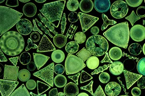
Background imageMicroscopic Collection: Diatoms - marine (x25)

Diatoms - marine (x25)
JC-567 Diatoms - marine (x25) John Clegg Please note that prints are for personal display purposes only and may not be reproduced in any way

Background imageMicroscopic Collection: Diatom - from marine plankton sample

Diatom - from marine plankton sample
SPH-530 Diatom - from marine plankton sample Santa Anna, California, USA Steve Hopkin Please note that prints are for personal display purposes only and may not be reproduced in any way

Background imageMicroscopic Collection: Illustration of a Flea C017 / 3435

Illustration of a Flea C017 / 3435
Plate from 18th century encyclopedia showing an illustration of a flea. Fleas are the insects forming the order Siphonaptera and are a vectors for various diseases

Background imageMicroscopic Collection: Under the Microscope / C18

Under the Microscope / C18
Animacules : microscopic creatures as seen under a microscope; the last two rows are human sperm

Background imageMicroscopic Collection: Macro Photograph: Patterns of Paenibacillus bacteria on petri dish - C type (chiral)

Macro Photograph: Patterns of Paenibacillus bacteria on petri dish - C type (chiral)
LRDS-218 Macro Photograph: Patterns of Paenibacillus bacteria on petri dish - C type (chiral) magnification x25 (A4 size: 29.7 cm width)

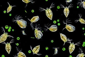
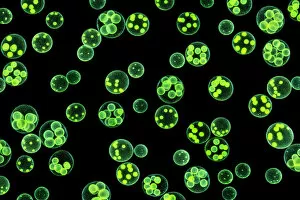
Background imageMicroscopic Collection: Water fleas (Daphnia sp. ) and a green algae (Volvox aureus) in water from a garden pond

Water fleas (Daphnia sp. ) and a green algae (Volvox aureus) in water from a garden pond
Water fleas (Daphnia sp.) and a green algae (Volvox aureus) in water from a garden pond. Derbyshire, UK. September. Digital composite

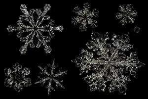
Background imageMicroscopic Collection: Different Snowflakes showing range in size and pattern, magnified under microscope

Different Snowflakes showing range in size and pattern, magnified under microscope, from Lilehammer, Norway. Digital composite

Background imageMicroscopic Collection: Chlorophyte or green alga (Volvox aureus) in pond water. UK. The larger spheres are approximately 0

Chlorophyte or green alga (Volvox aureus) in pond water. UK. The larger spheres are approximately 0.5mm in diameter. Digital composite image

Background imageMicroscopic Collection: Picture No. 11675528

Picture No. 11675528
Mouse Embryo at Eight Cell Stage. Date:

Background imageMicroscopic Collection: Picture No. 11675527

Picture No. 11675527
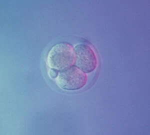
Mouse Embryo at Four Cell Stage & a Polar Body. Date:

Background imageMicroscopic Collection: Picture No. 11675526

Picture No. 11675526
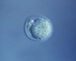
Mouse Embryo at Two Cell Stage & a Polar Body. Date:

Background imageMicroscopic Collection: Picture No. 11675525

Picture No. 11675525
Mouse Embryo at Single Cell Stage and Polar Body. Date:

Background imageMicroscopic Collection: Picture No. 11675522

Picture No. 11675522
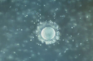
A human Ovum surrounded by Sperm - being fertilized. Date:

Background imageMicroscopic Collection: Picture No. 11675478

Picture No. 11675478
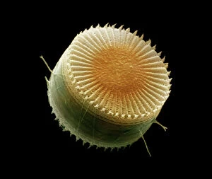
Scanning Electron Micrograph (SEM): Freshwater Diatom, Cyclotella meneghiniana. Date:

Background imageMicroscopic Collection: Picture No. 11675613

Picture No. 11675613
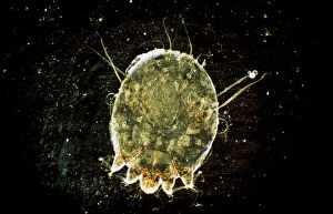
Dark Field Light Micrograph: Scabies Mite. Date:

Background imageMicroscopic Collection: Picture No. 11675612

Picture No. 11675612
Scanning Electron Micrograph (SEM): Human White and Red Blood Cells. Date:

Background imageMicroscopic Collection: Picture No. 11675599

Picture No. 11675599
Dark Field Light Micrograph: Lancet Liver Fluke. Date:

Background imageMicroscopic Collection: Picture No. 10876997

Picture No. 10876997
Scanning Electron Micrograph (SEM): Human Skin with Hair Follicles Date:

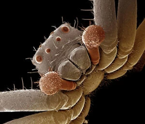
Background imageMicroscopic Collection: Scanning Electron Micrograph (SEM): Crab Spider, Magnification x 140 (A4 size: 29. 7 cm width)

Scanning Electron Micrograph (SEM): Crab Spider, Magnification x 140 (A4 size: 29. 7 cm width)
LRDS-120 Crab Spider Scanning Electron Micrograph (SEM) Xysticus sp. Magnification x 140 (A4 size: 29.7 cm width) Coloured by hand to enhance natural features

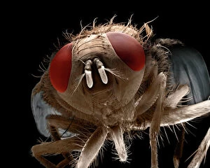
Background imageMicroscopic Collection: Scanning Electron Micrograph (SEM): House Fly, Magnification x 40 (A4 size: 29. 7 cm width)

Scanning Electron Micrograph (SEM): House Fly, Magnification x 40 (A4 size: 29. 7 cm width)
LRDS-72 House Fly Scanning Electron Micrograph (SEM) Musca domestica Magnification x 40 (A4 size: 29.7 cm width) Coloured by hand to enhance natural features

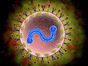
Background imageMicroscopic Collection: Microscopic view of human respiratory syncytial virus

Microscopic view of human respiratory syncytial virus (RSV). RSV causes respiratory tract infection of the lungs and breathing passages

Background imageMicroscopic Collection: Sugar crystals, ordinary table sugar, photomicrography

Sugar crystals, ordinary table sugar, photomicrography

Background imageMicroscopic Collection: Light Micrograph (LM): Protozoans: Kidney shaped ciliate surrounded by Euglena sp

Light Micrograph (LM): Protozoans: Kidney shaped ciliate surrounded by Euglena sp
LRDS-316 Light Micrograph (LM): Protozoans: Kidney shaped ciliate surrounded by Euglena sp. Magnification x 900 (when printed A4, 29.7 cm wide) Tillina sp

Background imageMicroscopic Collection: Picture No. 11675582

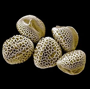
Picture No. 11675582
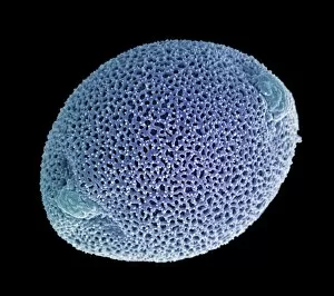
Scanning Electron micrograph (SEM): Lily Pollen grains. Date:

Background imageMicroscopic Collection: Picture No. 11675585

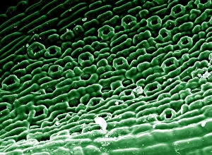
Picture No. 11675585
Scanning Electron micrograph (SEM)showing stomata on a Yew Leaf. Date:

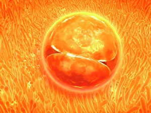
Background imageMicroscopic Collection: Embryo development 24-36 hours after fertilization

Embryo development 24-36 hours after fertilization

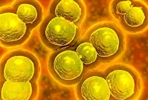
Background imageMicroscopic Collection: Microscopic view of chlamydia

Microscopic view of chlamydia. Chlamydia is a common sexually transmitted disease (STD) caused by a bacterium

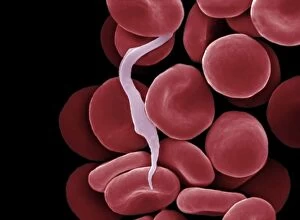
Background imageMicroscopic Collection: Picture No. 10873704

Picture No. 10873704
Sleeping Sickness Parasite in red blood cells (Trypanosoma sp. ) Date:

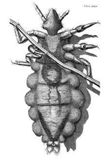
Background imageMicroscopic Collection: HOOKE: LOUSE, 1665. A louse, as seen by Robert Hooke with his microscope

HOOKE: LOUSE, 1665. A louse, as seen by Robert Hooke with his microscope. Copper engraving from Hookes Micrographia, 1665

Background imageMicroscopic Collection: Dinoflagellate plankton, SEM

Dinoflagellate plankton, SEM
Dinoflagellate plankton. Coloured scanning electron micrograph (SEM) of the calcareous (calcium-based) test (shell) of a dinoflagellate Calciodinellum sp

Background imageMicroscopic Collection: Macro Photograph: Patterns of Paenibacillus bacteria on petri dish - T type

Macro Photograph: Patterns of Paenibacillus bacteria on petri dish - T type (tip-splitting morphotype)
LRDS-232 Macro Photograph: Patterns of Paenibacillus bacteria on petri dish - T type (tip-splitting morphotype) magnification x5.6 (A4 size)

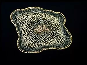
Background imageMicroscopic Collection: Light Micrograph (LM): A transverse section of a stem of Whisk Fern (Psilotum nudum)

Light Micrograph (LM): A transverse section of a stem of Whisk Fern (Psilotum nudum); Magnification x18 (on 10)
LRDS-167 Light Micrograph (LM): A transverse section of a stem of Whisk Fern (Psilotum nudum) Magnification x18 (on 10.5 cm width print) Psilotum nudum David Spears (Last Refuge)

Background imageMicroscopic Collection: False-coloured SEM (Scanning Electron Micrograph) of a Tardigrade (Tardigrada) amongst moss

False-coloured SEM (Scanning Electron Micrograph) of a Tardigrade (Tardigrada) amongst moss, from Peak District National Park, Derbyshire, UK. This tardigrade is approximately 0.1 millimetres long

Background imageMicroscopic Collection: Dinoflagellates, SEM

Dinoflagellates, SEM
Dinoflagellates. Coloured scanning electron micrograph (SEM) of Prorocentrum dinoflagellates (round) and Ceratium dinoflagellates (branched). Dinoflagellates are unicellular protozoans

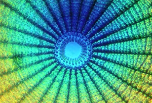
Background imageMicroscopic Collection: Diatom cell wall, SEM

Diatom cell wall, SEM
Diatom cell wall. Coloured scanning electron micrograph (SEM) of the pattern of small holes, called striae, in the cell wall of a diatom alga. This is a Coscinodiscus sp. diatom

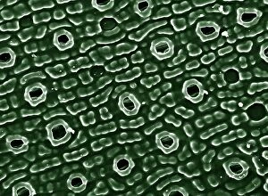
Background imageMicroscopic Collection: Picture No. 11675628

Picture No. 11675628
Scanning Electron Micrograph (SEM): Stomata of Yew Leaf. Date:

Background imageMicroscopic Collection: Microscopic Marine Life

Microscopic Marine Life
Various forms of microscopic marine life, described by Sibly as Animalcules Date: 1794

Background imageMicroscopic Collection: Scanning Electron Micrograph (SEM): Mosquito, Female; Magnification x 55 (A4 size: 29. 7 cm width)

Scanning Electron Micrograph (SEM): Mosquito, Female; Magnification x 55 (A4 size: 29. 7 cm width)
LRDS-83 Mosquito, Female Scanning Electron Micrograph (SEM) Anopheles sp. Magnification x 55 (A4 size: 29.7 cm width) Coloured by hand to enhance natural features

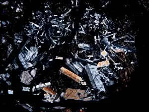
Background imageMicroscopic Collection: Microscope image of the Pasamonte eucrite

Microscope image of the Pasamonte eucrite
Microscopic image of the Pasamonte eucrite showing a basaltic texture. Field of view is 2.5mm across

Background imageMicroscopic Collection: Focus stacked, inverted image of Diatoms on a microscope slide

Focus stacked, inverted image of Diatoms on a microscope slide. Diatoms are single-celled algae which produce approximately 25% of the oxygen we breathe as well as being responsible for approximately

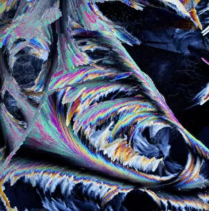
Background imageMicroscopic Collection: Sheet ice crystals viewed by polarised light

Sheet ice crystals viewed by polarised light

Background imageMicroscopic Collection: Specimen XII

Specimen XII
Amy Louise Baker


All Professionally Made to Order for Quick Shipping

"Exploring the Microscopic World: A Glimpse into Nature's Hidden Wonders" Step into a realm unseen by the naked eye, where tiny creatures and intricate structures come to life. In Picture No. 10851646, we are captivated by the Scanning Electron Micrograph (SEM) of a Praying Mantis, magnified x30. Its delicate features and mesmerizing details reveal a world within our reach. Moving on to another fascinating creature, the Tardigrade or 'Water Bear', magnified x1250 in an SEM image. This resilient micro-animal showcases its remarkable adaptability under extreme conditions. It reminds us that even in the tiniest forms of life, strength can be found. The Fruit Fly takes center stage next with an SEM image at x300 magnification. Despite its minuscule size, this common insect holds secrets waiting to be discovered under closer inspection. Its intricate body structure hints at its role in nature's grand tapestry. Diatoms from marine plankton samples grace our vision next - their elegant shapes and patterns resembling miniature works of art created by nature itself. These microscopic organisms play a vital role in Earth's ecosystems as primary producers. An illustration of a Flea C017/3435 introduces us to yet another hidden world beneath our feet - one filled with parasites and symbiotic relationships that shape entire ecosystems on both macro and micro scales. Diving deeper into marine diatoms at x25 magnification reveals their stunning beauty – intricate designs etched onto tiny shells floating through vast oceans, silently contributing to Earth's biodiversity. Anton van Leeuwenhoek's observations of Animalcules circa 1795 remind us of humanity's earliest encounters with microscopic lifeforms – pioneering discoveries that forever changed our understanding of biology and paved the way for modern scientific advancements. A Macro Photograph unveils Paenibacillus bacteria colonies thriving amidst laboratory-imposed stresses, showcasing their ability to adapt and survive in challenging environments.