Acrylic Blox : Hair shaft and skin, SEM


![]()
Mounted Prints from Science Photo Library
Hair shaft and skin, SEM
Hair shaft and skin. Coloured scanning electron micrograph (SEM) of a hair shaft (dark brown, left) growing from the surface of human skin. A hair shaft grows from a hair follicle (not seen) below the surface of the skin. Hair is made up of a fibrous protein called keratin. The outermost skin layer, the stratum corneum, also consists of keratinized dead cells (light brown) that are here detaching from the body. The squamous (flattened) cells that make up the stratum corneum arise from the lower, living layers of skin. Magnification: x167 at 6x7cm size
Science Photo Library features Science and Medical images including photos and illustrations
Media ID 6455913
© SUSUMU NISHINAGA/SCIENCE PHOTO LIBRARY
Cuticle Dead Epidermal Epidermis Follicle Hair Keratin Keratinized Scale Scaly Shaft Skin Squamous Stratum Corneum Surface Cells
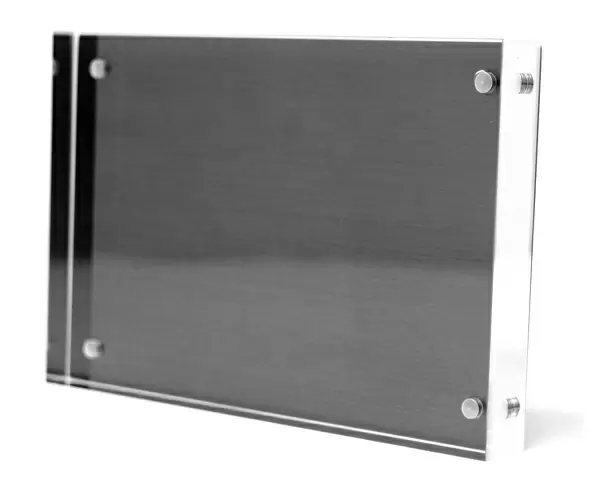
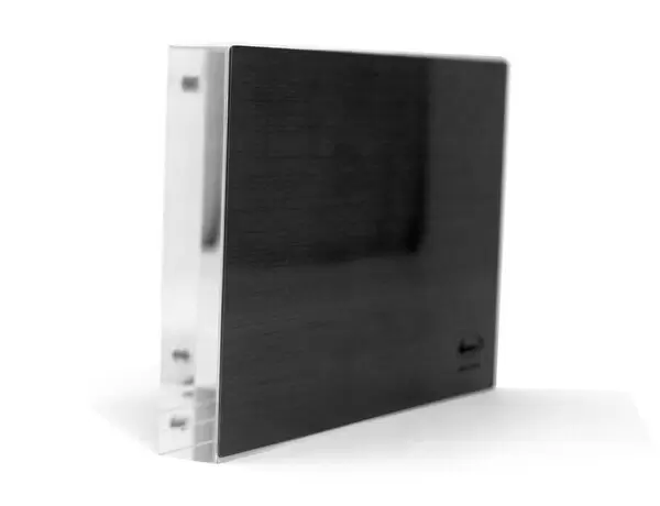
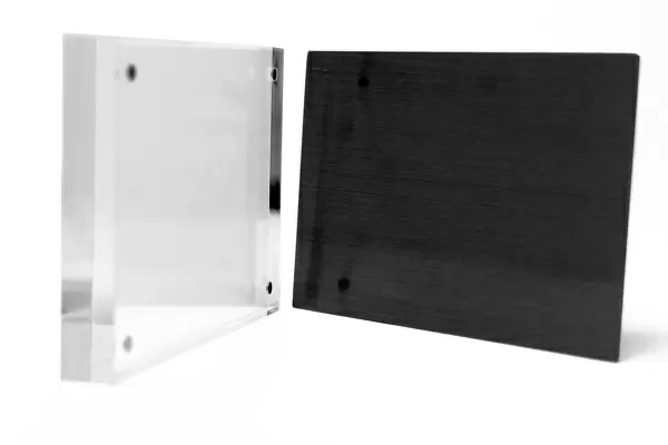
8"x6" (20x15cm) Acrylic Blox
Your photographic print is held in place by magnets and a micro thin sheet of metal covering the back of a 20mm piece of clear acrylic. Your print is held in place with magnets so can easily be replaced if needed.
Streamlined, one sided modern and attractive table top print
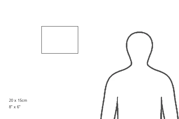
Estimated Product Size is 20.3cm x 15.2cm (8" x 6")
These are individually made so all sizes are approximate
Artwork printed orientated as per the preview above, with landscape (horizontal) orientation to match the source image.
EDITORS COMMENTS
This print showcases the intricate details of a hair shaft and the surrounding skin, captured using a scanning electron microscope (SEM). The dark brown hair shaft is seen emerging from the surface of human skin, originating from a hair follicle beneath. Composed primarily of keratin, a fibrous protein, this healthy strand epitomizes the natural beauty and resilience of our body's integumentary system. The image also reveals the outermost layer of skin known as the stratum corneum. In shades of light brown, these keratinized dead cells are shown detaching from the body, highlighting their role in maintaining our epidermal health. Interestingly, these squamous or flattened cells originate from deeper layers of living skin. With a magnification level set at x167 for this 6x7cm-sized print, every minute detail becomes visible to us. This photograph not only offers an insight into our anatomy but also serves as a reminder of how intricately designed our bodies are. Captured by Science Photo Library through advanced SEM technology, this image represents both scientific exploration and artistic appreciation for the wonders found within ourselves. It invites viewers to marvel at nature's complexity while contemplating their own unique composition on microscopic levels.
MADE IN THE UK
Safe Shipping with 30 Day Money Back Guarantee
FREE PERSONALISATION*
We are proud to offer a range of customisation features including Personalised Captions, Color Filters and Picture Zoom Tools
SECURE PAYMENTS
We happily accept a wide range of payment options so you can pay for the things you need in the way that is most convenient for you
* Options may vary by product and licensing agreement. Zoomed Pictures can be adjusted in the Basket.