
Squamous Collection
"Squamous: Exploring the Intricate Beauty of Skin Surface and Hair Structures" Delving beneath the surface


All Professionally Made to Order for Quick Shipping
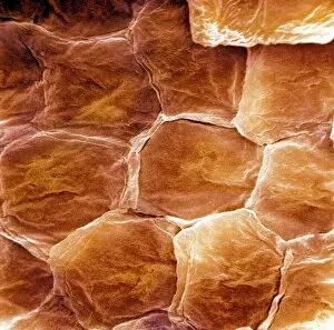
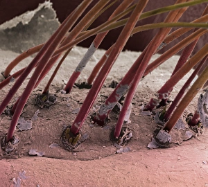
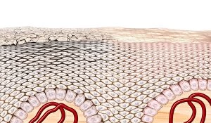
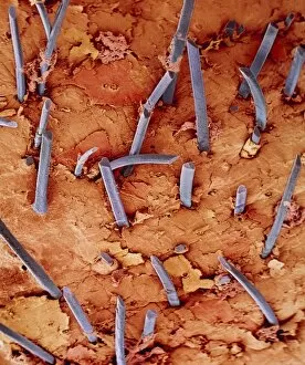
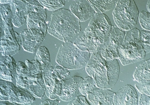
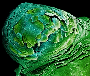
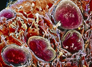
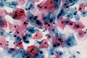
"Squamous: Exploring the Intricate Beauty of Skin Surface and Hair Structures" Delving beneath the surface, scanning electron microscopy (SEM) unravels the enigmatic world cells that make up our skin. From intricate patterns to delicate textures, each image reveals a fascinating story. In one captivating capture, we witness the tubercular syphilis symptoms etched on the body's canvas. A reminder of both the fragility and resilience of human skin, it serves as a testament to our ability to endure and heal. Moving closer still, SEM unveils eyelash hairs in all their mesmerizing glory. Each strand meticulously crafted by nature, they frame our eyes with elegance and grace - an exquisite detail often overlooked. Artwork C016/7541 takes us on an artistic journey through skin structure. Brushstrokes mimic its complexity, showcasing layers upon layers that protect us from external forces while allowing for vital exchanges within. Further exploration leads us into the depths of hair shafts intertwined with skin. SEM exposes eyebrow hairs in remarkable detail - tiny arches that define expression and add character to every face they adorn. Zooming even further down reveals individual skin cells under scrutiny. Their unique shapes and arrangements highlight their role as guardians against environmental stressors while maintaining harmony within. Back on familiar territory, SEM captures stubble hair emerging from pores like miniature forests reclaiming forgotten lands. A symbol of growth and transformation reminding us that change is constant in life's tapestry. Returning once more to examine every nook and cranny of our outermost layer, SEM uncovers secrets hidden within eyelash hairs' surfaces; a testament to nature's artistry at its finest - perfection found in imperfection. As we conclude this visual expedition through squamous wonders captured by SEM lenses, we are left awestruck by the sheer beauty residing just below what meets the eye.