Fraud Blocker Skip to main content

Stratum Corneum Collection

The stratum corneum, the outermost layer of the skin, is a fascinating subject to explore

Background imageStratum Corneum Collection: SEM of section through human skin

SEM of section through human skin
Skin. Coloured scanning electron micrograph of a section through the human skin. The uppermost layers of the skin (pink and red) make up the epidermis

Background imageStratum Corneum Collection: Skin layers, SEM

Skin layers, SEM

Background imageStratum Corneum Collection: Skin surface, SEM

Skin surface, SEM
Skin surface. Coloured scanning electron micrograph (SEM) of squamous cells on the surface of the skin. These are flat, keratinised

Background imageStratum Corneum Collection: Anatomy of the human skin

Anatomy of the human skin

Background imageStratum Corneum Collection: Diagram showing anatomy of human skin

Diagram showing anatomy of human skin

Background imageStratum Corneum Collection: Detailed cutaway diagram of human skin

Detailed cutaway diagram of human skin

Background imageStratum Corneum Collection: Different types of acne, non-inflammatory and inflammatory

Different types of acne, non-inflammatory and inflammatory

Background imageStratum Corneum Collection: Conceptual image of the layers of human skin

Conceptual image of the layers of human skin

Background imageStratum Corneum Collection: Eyelash hairs, SEM

Eyelash hairs, SEM
Eyelash hairs and skin. Coloured scanning electron micrograph (SEM) of eyelash hairs growing from the surface of human skin

Background imageStratum Corneum Collection: Cross section illustration of structure of human skin

Cross section illustration of structure of human skin

Background imageStratum Corneum Collection: Illustration of an atypical growth on the skin that could be a sign of skin cancer

Illustration of an atypical growth on the skin that could be a sign of skin cancer
Illustration of an atypical growth on the skin that could be a warning sign of skin cancer. Sudden change in the appearance, shape, color, size

Background imageStratum Corneum Collection: Human skin section, light micrograph

Human skin section, light micrograph
Human skin. Light micrograph of a section through healthy human skin. The outer surface of the skin is at top. The uppermost surface is the epidermis

Background imageStratum Corneum Collection: Human skin, SEM

Human skin, SEM
Human skin. Coloured scanning electron micrograph (SEM) of the surface layer of the human skin, the epidermis. The outer layer of the epidermis (the stratum corneum)

Background imageStratum Corneum Collection: Human skin section, light micrograph P710 / 0472

Human skin section, light micrograph P710 / 0472
Human skin. Light micrograph of a section through healthy human skin. The outer surface of the skin is at top. The uppermost surface is the epidermis

Background imageStratum Corneum Collection: Skin structure, artwork C016 / 7541

Skin structure, artwork C016 / 7541
Skin structure. Computer artwork showing the different layers and structure of human skin. The upper layer (centre) is the epidermis, with the dermis below

Background imageStratum Corneum Collection: Human Skin Cells (SEM)

Human Skin Cells (SEM)
Human Skin Cells. Scanning electron micrograph (SEM) of the surface layer of the human skin, the epidermis. The outer layer of the epidermis (stratum corneum)

Background imageStratum Corneum Collection: Skin anatomy, artwork

Skin anatomy, artwork
Skin anatomy. Artwork of a section through human skin, showing the flattening of skin cells as they rise to the dead keratinised surface layer (top) from the living layer below (the dermis)

Background imageStratum Corneum Collection: Hair shaft and skin, SEM

Hair shaft and skin, SEM
Hair shaft and skin. Coloured scanning electron micrograph (SEM) of a hair shaft (dark brown, left) growing from the surface of human skin

Background imageStratum Corneum Collection: Skin section, SEM

Skin section, SEM
Skin section. Coloured scanning electron micrograph (SEM) of a section through human skin. A hair (red) is protruding through the surface

Background imageStratum Corneum Collection: Eyebrow hair, SEM

Eyebrow hair, SEM
Eyebrow hair and skin. Coloured scanning electron micrograph (SEM) of eyebrow hair growing from the surface of human skin

Background imageStratum Corneum Collection: Coloured SEM of a section through skin

Coloured SEM of a section through skin
Skin. Coloured scanning electron micrograph of a section through human skin. The uppermost skin layer is the epidermis (orange & yellow)

Background imageStratum Corneum Collection: Skin layers, light micrograph

Skin layers, light micrograph
Skin layers. Coloured light micrograph of a section through human skin layers. The top layer is the stratum corneum (flaky, orange), a cornified layer of the epidermis that is composed of flattened

Background imageStratum Corneum Collection: Stubble hair, SEM

Stubble hair, SEM
Stubble hair and skin. Coloured scanning electron micrograph (SEM) of stubble hair growing from the surface of human skin

Background imageStratum Corneum Collection: Coloured SEM of a section through skin layers

Coloured SEM of a section through skin layers
Skin. Coloured scanning electron micrograph (SEM) of a section through human skin. At top is the stratum corneum (green) of the epidermis, a cornified layer composed of flattened

Background imageStratum Corneum Collection: Hair follicle, SEM

Hair follicle, SEM
Hair follicle. Coloured scanning electron micrograph (SEM) of an empty hair follicle (centre) among squamous cells on the surface of the skin

Background imageStratum Corneum Collection: Skin cells, SEM

Skin cells, SEM
Skin cells. Coloured scanning electron micrograph (SEM) of squamous cells from the surface of the skin. These are flat, keratinised

Background imageStratum Corneum Collection: Skin cell, SEM

Skin cell, SEM
Skin cell. Coloured scanning electron micrograph (SEM) of a squamous cell on the surface of the skin. This is a flat, keratinised, dead cell

Background imageStratum Corneum Collection: Human skin surface, SEM

Human skin surface, SEM
Human skin surface, coloured scanning electron micrograph (SEM). Ridges and loose squamous cells on the surface of the skin


All Professionally Made to Order for Quick Shipping

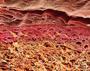
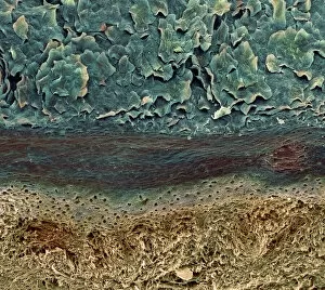
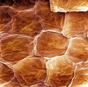
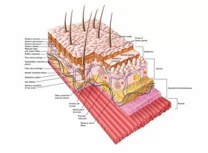
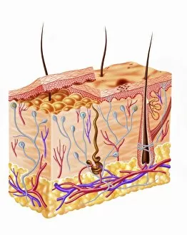
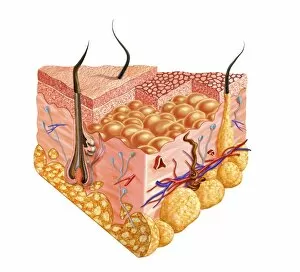
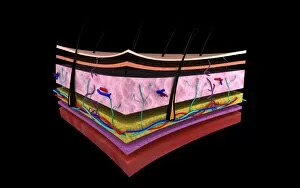
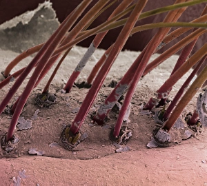
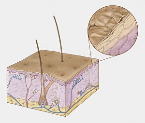
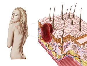
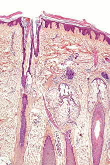
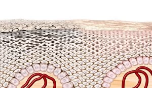
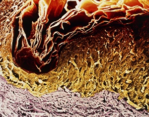
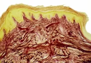
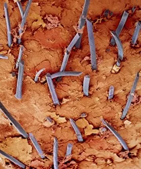
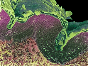
The stratum corneum, the outermost layer of the skin, is a fascinating subject to explore. Through scanning electron microscopy (SEM) of sections through human skin, we can witness its intricate structure and composition. This SEM image reveals the various layers that make up our skin, with the stratum corneum acting as a protective barrier against external threats. Another captivating SEM image showcases the surface of our skin in great detail. The texture and complexity of this layer are truly remarkable when observed at such high magnification. Understanding the anatomy of human skin is crucial for comprehending its functions and potential issues. From different types of acne like non-inflammatory and inflammatory forms to conceptual images depicting each layer's significance, these visuals provide valuable insights into dermatological concerns. Not limited to just facial features, SEM also allows us to examine other parts like eyelash hairs. These microscopic structures play an essential role in protecting our eyes from dust particles and debris. A cross-section illustration further elucidates the structure of human skin by highlighting its distinct layers. This biomedical depiction helps us grasp how each component contributes to overall health and well-being. However, it's important not only to appreciate but also be aware of potential risks associated with our largest organ. An illustration showcasing an atypical growth on the skin serves as a reminder that vigilance is necessary in detecting signs that could indicate conditions such as skin cancer.