
Keratin Collection
"Exploring the Fascinating World of Keratin: From Skin Surfaces to Exotic Creatures" Delving into the intricate world of keratin


All Professionally Made to Order for Quick Shipping
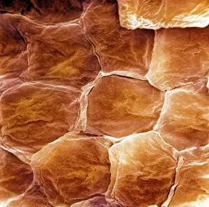
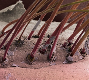
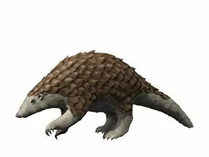
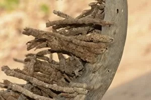
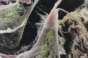
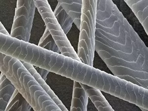
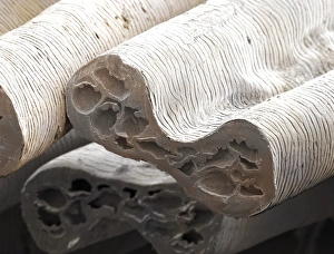
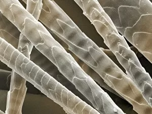
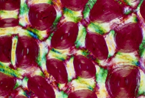
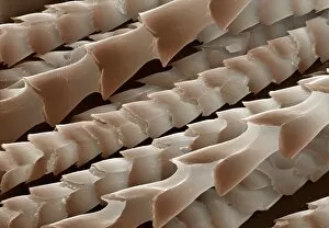
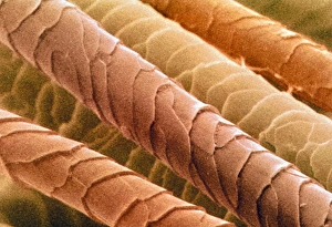
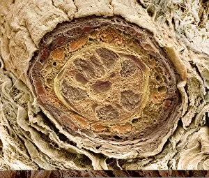
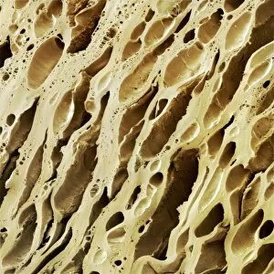
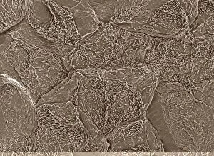
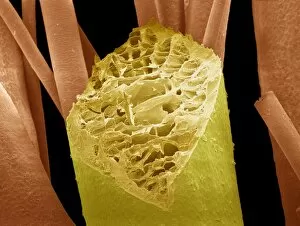
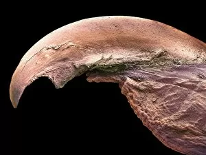
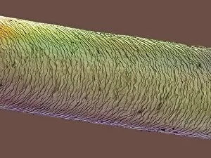
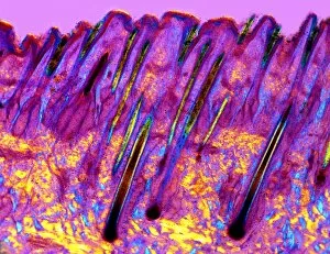
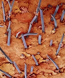
"Exploring the Fascinating World of Keratin: From Skin Surfaces to Exotic Creatures" Delving into the intricate world of keratin, a protein that plays a vital role in various aspects of life. Revealing the hidden beauty beneath our skin surface through scanning electron microscopy (SEM). Shedding light on nature's marvels with an SEM image capturing the delicate claw of a cat. Bathed in warm light, a captivating portrait captures the majestic pronghorn antelope buck in Yellowstone National Park, USA - its horns composed of sturdy keratin. Peering closely at eyelash hairs under SEM reveals their astonishing structure and composition. Journeying back in time to Germany's Eocene epoch, meet Eomanis waldi - a pangolin species whose scales are made up of keratinized material. Witnessing an extraordinary sight as horn moth larval faecal towers erupt from its horn - one of few creatures that feed on keratin-rich substances. Unveiling the microscopic wonders within tongues, where keratin covers the apex of papillae - providing essential functions for taste and texture perception. Exploring different animal hairs under colored SEM: domestic cat hair showcases mesmerizing patterns (colored SEM Z934 / 0486). Admiring red deer hair under colored SEM (Z952 / 0094), highlighting its unique characteristics and resilience thanks to keratin fibers. Discovering nature's diversity through grey squirrel hair seen under colored SEM - each strand revealing intricate details about this adaptable creature's fur coat composition. Examining wild rabbit hair under colored SEM exposes fascinating textures and structures shaped by resilient strands rich in keratin proteins. 13. Traveling across continents to witness camel hair like never before – vibrant colors reveal intricate patterns found within these durable fibers (colored SEM Z949 / 0010). 14.