Home > Popular Themes > Human Body
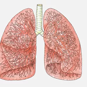
Respiratory nerves, 1844 artwork

![]()
Wall Art and Photo Gifts from Science Photo Library
Respiratory nerves, 1844 artwork
Respiratory nerves. These anatomical artworks are figures 2-3, plate 49, volume 3 (1844) of Traite complet de l anatomie de l homme (1831-1854). This 8-volume anatomy atlas was produced by the French physician and anatomist Jean-Baptiste Marc Bourgery (1797-1849). The illustrations were by Nicolas-Henri Jacob (1781-1871)
Science Photo Library features Science and Medical images including photos and illustrations
Media ID 6324951
© SCIENCE PHOTO LIBRARY
1844 Anatomical Artwork Anatomical Illustration Anatomy Atlas Blood Vessels Bronchi Bronchial Bronchus Chest Comprenant La Medecine Operatoire Dissected Dissection French Glossopharyngeal Nerve Hypoglossal Nerve Lung Lungs Nerve Nerves Neural Oesophagus Pulmonary Respiratory Thorax Traite Complet De Lanatomie De Lhomme Vagus Nerve Artery Nervous System Neurological Neurology Vein
EDITORS COMMENTS
This artwork, titled "Respiratory Nerves" is a remarkable piece from the 19th century. Created by French physician and anatomist Jean-Baptiste Marc Bourgery, in collaboration with illustrator Nicolas-Henri Jacob, this print is part of an eight-volume anatomy atlas called "Traite complet de l'anatomie de l'homme". The intricate illustration showcases the complexity of the respiratory system within the human body. It highlights various components such as veins, arteries, bronchi, and nerves that play crucial roles in our ability to breathe. With meticulous detail and precision, Bourgery and Jacob bring to life the inner workings of our thorax. The delicate interplay between blood vessels and neural pathways is beautifully depicted, providing a deeper understanding of how these systems function together harmoniously. This historical artwork not only serves as a testament to the advancements made in medical science during that era but also stands as a work of art in its own right. Its fusion of scientific accuracy with artistic finesse makes it both visually stunning and intellectually enlightening. As we gaze upon this masterpiece from Science Photo Library's collection, we are reminded of the immense intricacy hidden beneath our skin. It invites us to marvel at the wonders of human anatomy while paying homage to those who dedicated their lives to unraveling its mysteries.
MADE IN THE UK
Safe Shipping with 30 Day Money Back Guarantee
FREE PERSONALISATION*
We are proud to offer a range of customisation features including Personalised Captions, Color Filters and Picture Zoom Tools
SECURE PAYMENTS
We happily accept a wide range of payment options so you can pay for the things you need in the way that is most convenient for you
* Options may vary by product and licensing agreement. Zoomed Pictures can be adjusted in the Basket.