
Anatomical Artwork Collection
"Exploring the Intricacies of the Human Body: Anatomical Artwork Unveiled" Step into a world where art and science intertwine


All Professionally Made to Order for Quick Shipping
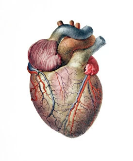
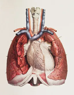
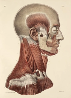
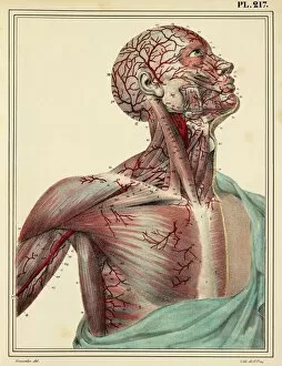
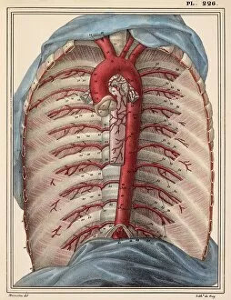
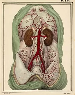
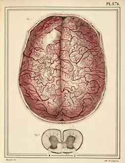
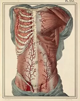
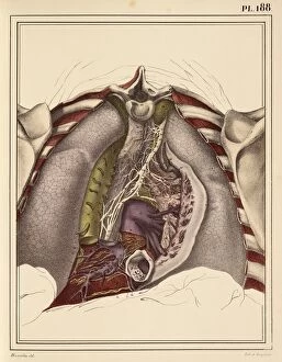
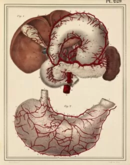
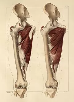
"Exploring the Intricacies of the Human Body: Anatomical Artwork Unveiled" Step into a world where art and science intertwine, revealing the hidden wonders within our bodies. This captivating collection takes us on a journey through various systems, showcasing their complexity and beauty. Intricate strokes bring to life the delicate network of blood vessels that nourish our brain, while vibrant hues depict the pulsating rhythm of our beating heart. A masterpiece from 1825 unveils an astonishing depiction of male groin arteries, reminding us of the intricate web that sustains life. Moving further down, we encounter a mesmerizing portrayal of the heart and lungs working in perfect harmony; each brushstroke capturing their vital dance. The attention then shifts to head and neck muscles in another remarkable piece from 1831 - every contour meticulously crafted to showcase their strength and grace. Delving deeper into this artistic exploration, we are transported to 1825 with an awe-inspiring artwork unveiling head and chest arteries. Here, we witness nature's own masterpiece as these lifelines branch out like rivers flowing through a vast landscape. The focus now turns towards face and neck muscles portrayed in yet another stunning work from 1831. Each muscle is meticulously etched onto canvas, highlighting their role in expressions both subtle and profound – truly capturing human emotion frozen in time. As we continue this visual expedition through time, cervical spinal nerves take center stage in a breathtaking creation dating back to 1844. Every nerve intricately detailed reminds us how interconnected our body truly is – a symphony orchestrated by nature itself. Our hands hold secrets too; revealed through an exquisite artwork from 1831 depicting hand muscle anatomy. These intricate structures enable us to create art ourselves – tools for expression that often go unnoticed until brought forth on canvas. Muscles come alive once more as we explore forearm anatomy showcased brilliantly in another piece from 1831.