
Anatomy Atlas Collection
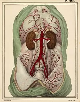
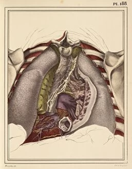
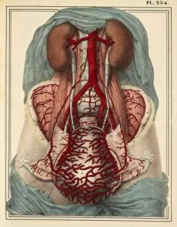
Step into the fascinating world of human anatomy with this captivating 1825 artwork, showcasing the intricate details of male groin arteries


All Professionally Made to Order for Quick Shipping
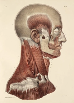
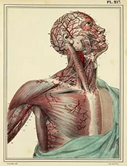
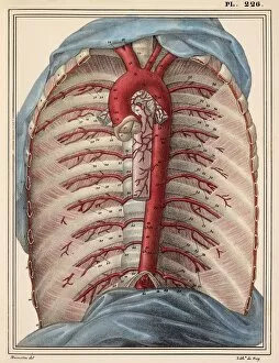
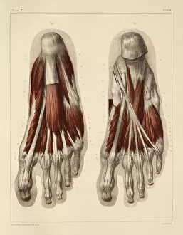
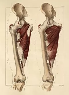
Step into the fascinating world of human anatomy with this captivating 1825 artwork, showcasing the intricate details of male groin arteries. Delve deeper into the complexity of our bodies as you explore the 1831 artwork depicting head and neck muscles, revealing a network of strength and flexibility. Transport yourself to another era as you uncover the hidden secrets within the head and chest arteries in an exquisite piece from 1825. Marvel at the precision captured in every stroke, bringing these vital pathways to life on paper. Embark on a journey through time with a stunning 1831 artwork highlighting face and neck muscles, illustrating their role in expression and movement. Witness how each muscle contributes to our unique identities. Discover the wonders of cervical spinal nerves through an extraordinary masterpiece from 1844. This detailed illustration offers insight into our body's communication system, unveiling its intricacies like never before. Immerse yourself in hand muscle anatomy depicted beautifully in an 1831 artwork that showcases both form and function. Appreciate how these small yet mighty structures enable us to perform delicate tasks with precision. Continue your exploration by studying muscles of the forearm meticulously portrayed in an awe-inspiring piece from 1831. Gain a newfound appreciation for their power and versatility as they facilitate countless movements throughout our daily lives. Uncover the strength within pelvic-femoral muscles through an evocative artwork dating back to 1831. Witness firsthand how these essential structures provide stability while allowing for graceful mobility. Marvel at armpit muscles showcased brilliantly in an enchanting piece from 1831, shedding light on their often overlooked significance within our musculoskeletal system. Delve into face and neck nerves immortalized in breathtaking detail by talented artists back in 1825. Understand how these intricate networks transmit information crucial for sensation, movement, and expression. Explore superficial back muscles brought vividly to life through mesmerizing brushstrokes captured flawlessly by skilled hands during the year 1831.