Home > Science > SEM
Plant cell, SEM

![]()
Wall Art and Photo Gifts from Science Photo Library
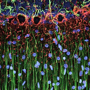
Plant cell, SEM
Plant cell. Coloured scanning electron micrograph (SEM) of a section through a plant cell, revealing its internal structure. The cell is encased in a cellulose, hemicellulose and pectin cell wall. Inside the cell wall are chloroplasts (dark green), the site of photosynthesis, and the nucleus (orange), which contains the cells genetic information. At the centre of the cell is a large vacuole, which maintains the cells shape, stores useful materials and digests the cells waste products
Science Photo Library features Science and Medical images including photos and illustrations
Media ID 6305699
© DR DAVID FURNESS, KEELE UNIVERSITY/SCIENCE PHOTO LIBRARY
Cell Biology Cell Wall Chloroplast Chloroplasts Cytology Eukaryote Eukaryotic Interior Nucleus Organelle Organelles Plant Cell Plastid Plastids Vacuole False Coloured Section Sectioned
EDITORS COMMENTS
This print showcases the intricate and vibrant world of a plant cell. Through the lens of a scanning electron microscope, we are granted access to its hidden interior structure, revealing an awe-inspiring complexity. Encased in a sturdy yet flexible cell wall composed of cellulose, hemicellulose, and pectin, this plant cell stands as a testament to nature's ingenuity. Within this protective barrier lies an array of essential components that sustain life itself. The chloroplasts take center stage with their dark green hue, serving as the powerhouses responsible for photosynthesis - the process by which plants convert sunlight into energy. Their presence highlights the remarkable ability of plants to harness light and transform it into sustenance. Nestled alongside these chloroplasts is the nucleus, represented here in vivid orange tones. This vital organelle houses the genetic information that defines every aspect of this living entity's existence. At the heart of this bustling cellular landscape resides a large vacuole - a multifunctional space that maintains shape while storing valuable materials and breaking down waste products. It serves as both protector and recycler within this microcosm. As we gaze upon this false-colored masterpiece captured by Science Photo Library's skilled photographers, we are reminded once again of nature's boundless beauty and complexity. This image not only celebrates botanical wonders but also invites us to marvel at life on its smallest scale – where even within something seemingly simple like a plant cell lies an entire universe waiting to be explored
MADE IN THE UK
Safe Shipping with 30 Day Money Back Guarantee
FREE PERSONALISATION*
We are proud to offer a range of customisation features including Personalised Captions, Color Filters and Picture Zoom Tools
SECURE PAYMENTS
We happily accept a wide range of payment options so you can pay for the things you need in the way that is most convenient for you
* Options may vary by product and licensing agreement. Zoomed Pictures can be adjusted in the Basket.