
Eukaryote Collection
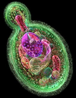
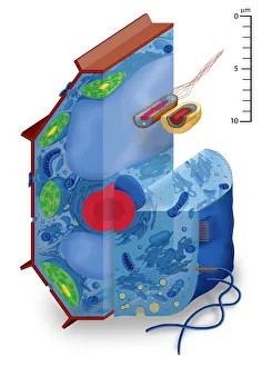
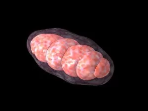
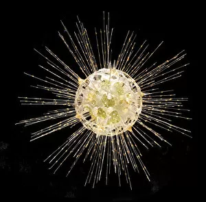
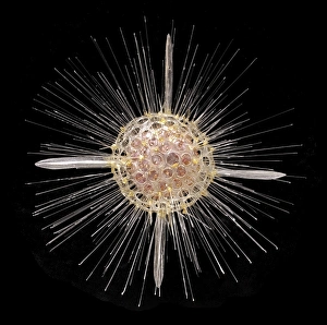
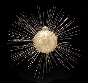
Eukaryotes, the diverse group of organisms that possess a true nucleus within their cells, encompass an array of fascinating life forms


All Professionally Made to Order for Quick Shipping
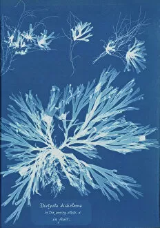
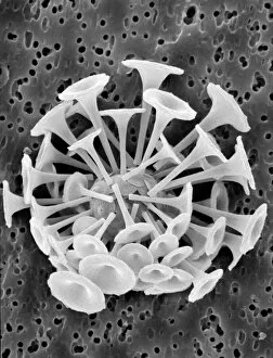
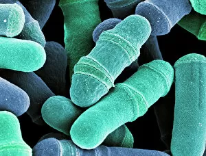
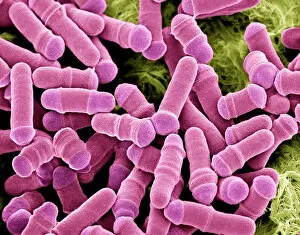
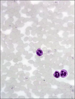
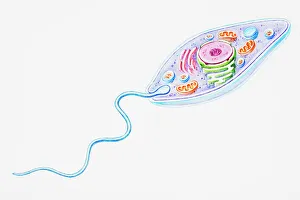
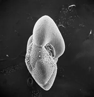
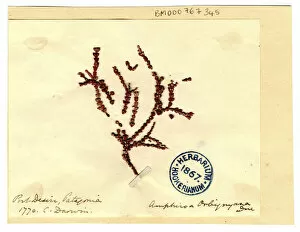
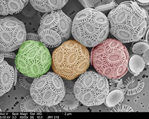
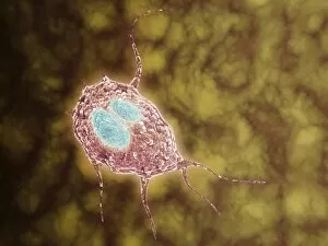
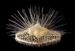
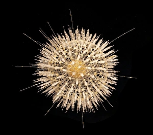
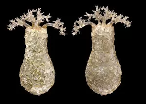
Eukaryotes, the diverse group of organisms that possess a true nucleus within their cells, encompass an array of fascinating life forms. From budding yeast cells to pressed seaweed specimens and coccolithophores like Discosphaera tubifera, eukaryotes showcase incredible diversity. Dictyota dichotoma and Fucus bulbosus, commonly known as kelp, exemplify the beauty and complexity found in eukaryotic cell types. These intricate structures are not limited to artwork; they exist in nature as well. Under the scanning electron microscope (SEM), dividing yeast cells reveal their remarkable process of reproduction. The marine world is teeming with eukaryotic wonders such as Fucus radiatus and Acanthophracta radiolarians. Their unique characteristics highlight the vastness of this kingdom's biodiversity. But it's not just multicellular organisms that fall under the umbrella of eukaryotes. Protozoa, single-celled organisms like Plasmodium sp. , exhibit complex behaviors while scavenging for particles or absorbing nutrients from their environment. Whether you're marveling at a budding yeast cell or examining pressed seaweed specimens C016/6127, exploring the realm of eukaryotes reveals a captivating tapestry woven by nature itself.