
Chloroplast Collection
Chloroplasts, the powerhouses of plant cells, are truly captivating structures. Within the cell types of a pea plant, these they can be found in abundance


All Professionally Made to Order for Quick Shipping
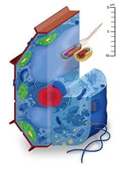
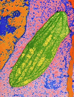
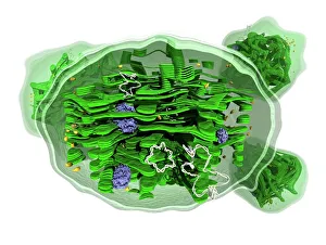
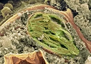
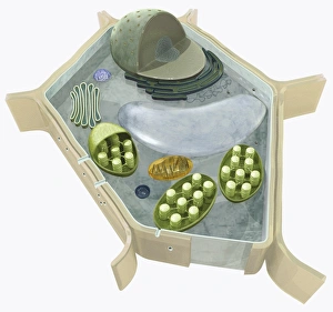
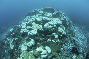
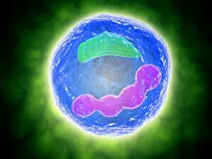
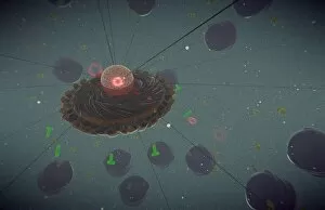
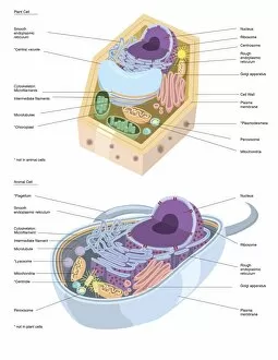
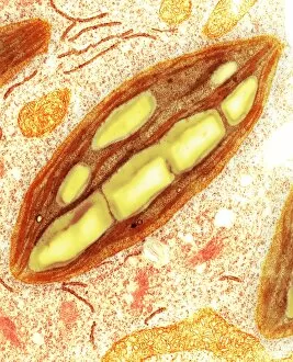
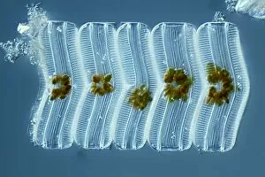
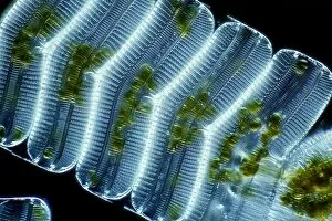
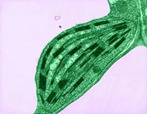
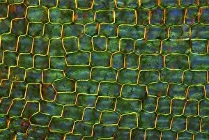
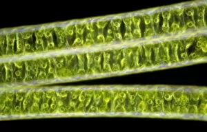
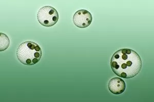
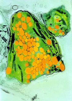
Chloroplasts, the powerhouses of plant cells, are truly captivating structures. Within the cell types of a pea plant, these they can be found in abundance. Their vibrant green color and intricate structure make them perfect subjects for artwork that showcases their beauty. When examining a chloroplast's structure closely, one can marvel at its complexity. With layers upon layers of membranes and stacks of thylakoids containing pigments like chlorophyll, it is no wonder they play a crucial role in photosynthesis. In an SEM image capturing the essence of a chloroplast, its detailed features become even more pronounced. The magnified view reveals the inner workings of this organelle - from grana to stroma - each contributing to the overall function. An illustration depicting the structure of a plant cell gives us insight into where exactly we can find these remarkable chloroplasts alongside other essential components such as the nucleus, nucleolus, ribosome, and endoplasmic reticulum. Together they form an intricate network that sustains life within plants. However, it is not just plants that rely on chloroplasts; coral colonies too depend on their symbiotic relationship with algae-containing chloroplasts called zooxanthellae. Sadly though, corals are beginning to bleach on reefs in Indonesia due to various environmental factors disrupting this delicate balance. As we witness corals bleaching before our eyes through images captured underwater off Indonesian shores or conceptual illustrations portraying this phenomenon's impact on marine ecosystems – it serves as a stark reminder of how interconnected our world truly is. Whether appreciating their artistic representation or understanding their vital role in sustaining life both above and below water surfaces – studying and acknowledging the significance of chloroplasts allows us to appreciate nature's incredible intricacies while also recognizing our responsibility towards preserving it for future generations.