Premium Framed Print > Animals > Mammals > Nesomyidae > Fat Mouse
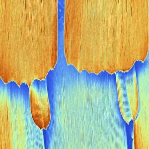
Premium Framed Print : TEM of endoplasmic reticulum in mammalian cell


![]()
Framed Photos from Science Photo Library
TEM of endoplasmic reticulum in mammalian cell
Rough endoplasmic reticulum. Transmission Electron Micrograph (TEM) of a section through a mammalian cell revealing rough endoplasmic reticulum (ER). ER is the system of membranes seen running across at upper image. Small granules attached to the membrane of the ER are ribosomes, the sites of protein synthesis. At lower right the large mass of the cell nucleus is seen, with dark areas of chromatin. ER originates from the nuclear membrane & divides the cell cytoplasm into a network of cavities called cisternae. Small vesicles bud from the ER to transport proteins and fats around the cell. Magnification: x66, 000 at 8x10 inch size
Science Photo Library features Science and Medical images including photos and illustrations
Media ID 6401483
© NIBSC/SCIENCE PHOTO LIBRARY
Cell Structure Cytology Endoplasmic Reticulum Protein Synthesis Ribosome Rough Rough Endoplasmic Reticulum Micro Biology
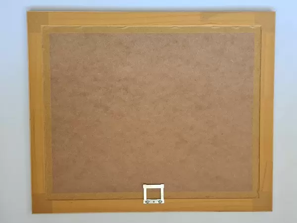
17"x15" (43x38cm) Premium Frame
FSC real wood frame with double mounted 10x8 print. Double mounted with white conservation mountboard. Frame moulding comprises stained composite natural wood veneers (Finger Jointed Pine) 39mm wide by 21mm thick. Archival quality Fujifilm CA photo paper mounted onto 1mm card. Overall outside dimensions are 17x15 inches (431x381mm). Rear features Framing tape to cover staples, 50mm Hanger plate, cork bumpers. Glazed with durable thick 2mm Acrylic to provide a virtually unbreakable glass-like finish. Acrylic Glass is far safer, more flexible and much lighter than typical mineral glass. Moreover, its higher translucency makes it a perfect carrier for photo prints. Acrylic allows a little more light to penetrate the surface than conventional glass and absorbs UV rays so that the image and the picture quality doesn't suffer under direct sunlight even after many years. Easily cleaned with a damp cloth. Please note that, to prevent the paper falling through the mount window and to prevent cropping of the original artwork, the visible print may be slightly smaller to allow the paper to be securely attached to the mount without any white edging showing and to match the aspect ratio of the original artwork.
FSC Real Wood Frame and Double Mounted with White Conservation Mountboard - Professionally Made and Ready to Hang
Estimated Image Size (if not cropped) is 19.1cm x 24.4cm (7.5" x 9.6")
Estimated Product Size is 38.1cm x 43.1cm (15" x 17")
These are individually made so all sizes are approximate
Artwork printed orientated as per the preview above, with portrait (vertical) orientation to match the source image.
FEATURES IN THESE COLLECTIONS
> Animals
> Mammals
> Nesomyidae
> Fat Mouse
> Animals
> Mammals
> Small Mammals
EDITORS COMMENTS
This print showcases the intricate details of a mammalian cell's endoplasmic reticulum (ER) as captured through a transmission electron microscope (TEM). The upper image reveals the rough ER, characterized by its system of membranes running across. Attached to these membranes are small granules known as ribosomes, which serve as vital sites for protein synthesis within the cell. In the lower right corner, we observe the prominent mass of the cell nucleus with dark areas representing chromatin. It is worth noting that the ER originates from the nuclear membrane and plays a crucial role in dividing the cytoplasm into interconnected cavities called cisternae. The complexity and functionality of this cellular structure become apparent when considering that small vesicles bud from the ER to facilitate transportation of proteins and fats throughout various regions of the cell. This process ensures efficient communication and distribution within this microscopic world. With a magnification level reaching an astonishing x66,000 at an 8x10 inch size, this TEM image offers us a glimpse into one aspect of biological microcosms. Science Photo Library has expertly captured this stunning visual representation without any commercial intent but rather with a focus on showcasing scientific marvels like this photograph for educational purposes in fields such as biology, cytology, and micro-biology.
MADE IN THE UK
Safe Shipping with 30 Day Money Back Guarantee
FREE PERSONALISATION*
We are proud to offer a range of customisation features including Personalised Captions, Color Filters and Picture Zoom Tools
SECURE PAYMENTS
We happily accept a wide range of payment options so you can pay for the things you need in the way that is most convenient for you
* Options may vary by product and licensing agreement. Zoomed Pictures can be adjusted in the Basket.