Premium Framed Print > Science > SEM
Premium Framed Print : False-colour SEM of embryo at the morula stage


![]()
Framed Photos from Science Photo Library
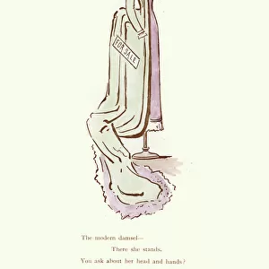
False-colour SEM of embryo at the morula stage
Embryo development. False-colour scanning electron micrograph of an embryo at the early stage known as the morula. The egg reaches this phase about 4 days after fertilisation after a series of mitotic divisions. At this stage about 12-16 cells are present and are surrounded by a thin glycoprotein layer, the zona pellucida, which was here removed. The inner cells of the morula will give rise to the tissues of the embryo while the outer cells, covered here by microvilli (tiny orange ridges), will form the placenta. The morula will implant into the uterus six days after fertilisation. Magnification: x645 at 6x7cm size. Magnification: x1005 at 4x5 inch size. This is a mouse morula
Science Photo Library features Science and Medical images including photos and illustrations
Media ID 6453919
© PROFESSORS P.M. MOTTA & J. VAN BLERKOM/ SCIENCE PHOTO LIBRARY
Cell Division Embryo Magnified Image Microscopic Photos Morula Subjects False Coloured
17"x15" (43x38cm) Premium Frame
FSC real wood frame with double mounted 10x8 print. Double mounted with white conservation mountboard. Frame moulding comprises stained composite natural wood veneers (Finger Jointed Pine) 39mm wide by 21mm thick. Archival quality Fujifilm CA photo paper mounted onto 1mm card. Overall outside dimensions are 17x15 inches (431x381mm). Rear features Framing tape to cover staples, 50mm Hanger plate, cork bumpers. Glazed with durable thick 2mm Acrylic to provide a virtually unbreakable glass-like finish. Acrylic Glass is far safer, more flexible and much lighter than typical mineral glass. Moreover, its higher translucency makes it a perfect carrier for photo prints. Acrylic allows a little more light to penetrate the surface than conventional glass and absorbs UV rays so that the image and the picture quality doesn't suffer under direct sunlight even after many years. Easily cleaned with a damp cloth. Please note that, to prevent the paper falling through the mount window and to prevent cropping of the original artwork, the visible print may be slightly smaller to allow the paper to be securely attached to the mount without any white edging showing and to match the aspect ratio of the original artwork.
FSC Real Wood Frame and Double Mounted with White Conservation Mountboard - Professionally Made and Ready to Hang
Estimated Image Size (if not cropped) is 21.3cm x 24.4cm (8.4" x 9.6")
Estimated Product Size is 38.1cm x 43.1cm (15" x 17")
These are individually made so all sizes are approximate
Artwork printed orientated as per the preview above, with portrait (vertical) orientation to match the source image.
FEATURES IN THESE COLLECTIONS
EDITORS COMMENTS
This print showcases the intricate details of an embryo at the morula stage, providing a fascinating glimpse into early stages of development. Through false-colour scanning electron microscopy, we are able to witness the remarkable process of cell division and differentiation. At this particular stage, approximately four days after fertilisation, the morula consists of 12-16 cells enveloped by a delicate glycoprotein layer called the zona pellucida. In this image, the zona pellucida has been meticulously removed to reveal both inner and outer cells. The inner cells hold immense potential as they will eventually give rise to various tissues within the developing embryo. The outer cells depicted here are adorned with microvilli – tiny orange ridges that provide additional surface area for nutrient absorption. These specialized structures indicate their crucial role in forming the placenta, which is vital for nourishing and supporting fetal growth during pregnancy. Through magnification at x645 (6x7cm size) or x1005 (4x5 inch size), we can truly appreciate the intricacies of embryonic development on a microscopic level. This specific image captures a mouse morula; however, it serves as an invaluable representation of early-stage embryo development across species. Science Photo Library presents this awe-inspiring photograph as part of its extensive collection featuring subjects like human body, embryo development, cell division, and microscopic photos.
MADE IN THE UK
Safe Shipping with 30 Day Money Back Guarantee
FREE PERSONALISATION*
We are proud to offer a range of customisation features including Personalised Captions, Color Filters and Picture Zoom Tools
SECURE PAYMENTS
We happily accept a wide range of payment options so you can pay for the things you need in the way that is most convenient for you
* Options may vary by product and licensing agreement. Zoomed Pictures can be adjusted in the Basket.