Framed Print > Science > SEM
Framed Print : False-colour SEM of embryo at the morula stage


![]()
Framed Photos from Science Photo Library
False-colour SEM of embryo at the morula stage
Embryo development. False-colour scanning electron micrograph of an embryo at the early stage known as the morula. The egg reaches this phase about 4 days after fertilisation after a series of mitotic divisions. At this stage about 12-16 cells are present and are surrounded by a thin glycoprotein layer, the zona pellucida, which was here removed. The inner cells of the morula will give rise to the tissues of the embryo while the outer cells, covered here by microvilli (tiny orange ridges), will form the placenta. The morula will implant into the uterus six days after fertilisation. Magnification: x645 at 6x7cm size. Magnification: x1005 at 4x5 inch size. This is a mouse morula
Science Photo Library features Science and Medical images including photos and illustrations
Media ID 6453919
© PROFESSORS P.M. MOTTA & J. VAN BLERKOM/ SCIENCE PHOTO LIBRARY
Cell Division Embryo Magnified Image Microscopic Photos Morula Subjects False Coloured
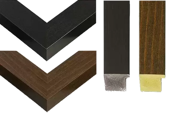
14"x12" (38x32cm) Modern Frame
Discover the wonders of embryonic development with our captivating Framed Prints from Media Storehouse. This particular print showcases a False-colour Scanning Electron Micrograph of an embryo at the morula stage, captured by Science Photo Library. At approximately 4 days after fertilization, the egg enters this early phase, marked by the formation of a compact mass of cells. Bring this stunning image into your home or office to inspire curiosity and ignite conversations about the marvels of science and life. Our high-quality framed prints are meticulously crafted to preserve the vivid colors and intricate details of the original image, ensuring a stunning addition to any space.
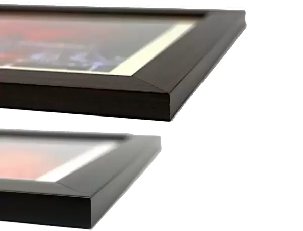
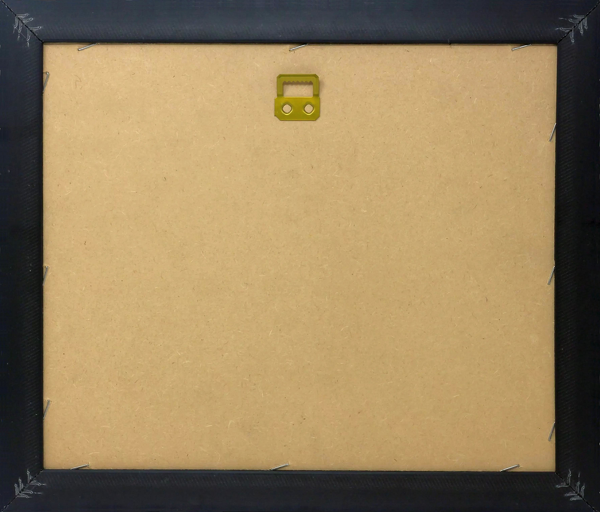
Wood effect frame, card mounted, 10x8 archival quality photo print. Overall outside dimensions 14x12 inches (38x32cm). Environmentally and ozone friendly, 40mm wide x 15mm Polycore® moulding has the look of real wood, is durable and light and easy to hang. Biodegradable and made with non-chlorinated gases (no toxic fumes) it is efficient; producing 100 tons of polystyrene can save 300 tons of trees! Prints are glazed with lightweight, shatterproof, optical clarity acrylic (providing the same general protection from the environment as glass). The back is stapled hardboard with a sawtooth hanger attached. Note: To minimise original artwork cropping, for optimum layout, and to ensure print is secure, the visible print may be marginally smaller
Contemporary Framed and Mounted Prints - Professionally Made and Ready to Hang
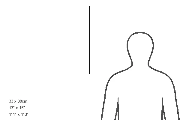
Estimated Image Size (if not cropped) is 21.3cm x 24.4cm (8.4" x 9.6")
Estimated Product Size is 32.5cm x 37.6cm (12.8" x 14.8")
These are individually made so all sizes are approximate
Artwork printed orientated as per the preview above, with portrait (vertical) orientation to match the source image.
FEATURES IN THESE COLLECTIONS
EDITORS COMMENTS
This print showcases the intricate details of an embryo at the morula stage, providing a fascinating glimpse into early stages of development. Through false-colour scanning electron microscopy, we are able to witness the remarkable process of cell division and differentiation. At this particular stage, approximately four days after fertilisation, the morula consists of 12-16 cells enveloped by a delicate glycoprotein layer called the zona pellucida. In this image, the zona pellucida has been meticulously removed to reveal both inner and outer cells. The inner cells hold immense potential as they will eventually give rise to various tissues within the developing embryo. The outer cells depicted here are adorned with microvilli – tiny orange ridges that provide additional surface area for nutrient absorption. These specialized structures indicate their crucial role in forming the placenta, which is vital for nourishing and supporting fetal growth during pregnancy. Through magnification at x645 (6x7cm size) or x1005 (4x5 inch size), we can truly appreciate the intricacies of embryonic development on a microscopic level. This specific image captures a mouse morula; however, it serves as an invaluable representation of early-stage embryo development across species. Science Photo Library presents this awe-inspiring photograph as part of its extensive collection featuring subjects like human body, embryo development, cell division, and microscopic photos.
MADE IN THE UK
Safe Shipping with 30 Day Money Back Guarantee
FREE PERSONALISATION*
We are proud to offer a range of customisation features including Personalised Captions, Color Filters and Picture Zoom Tools
SECURE PAYMENTS
We happily accept a wide range of payment options so you can pay for the things you need in the way that is most convenient for you
* Options may vary by product and licensing agreement. Zoomed Pictures can be adjusted in the Basket.