
Embryo Collection
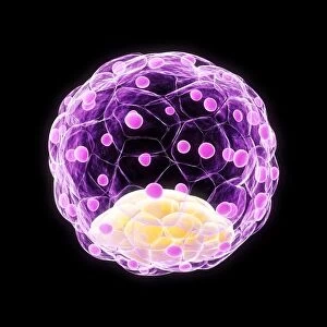
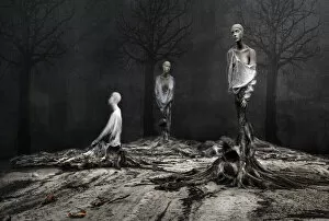
Embryo - a tiny marvel of life, the beginning of a new existence


All Professionally Made to Order for Quick Shipping
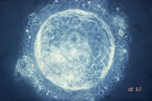
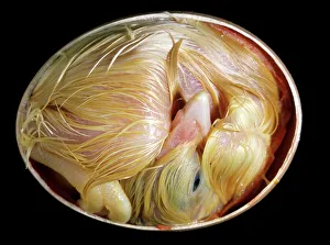
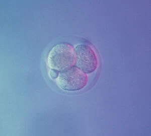
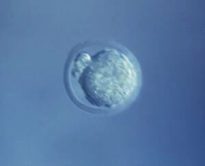
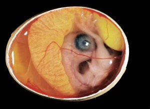
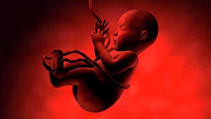
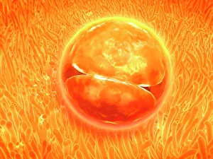
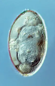
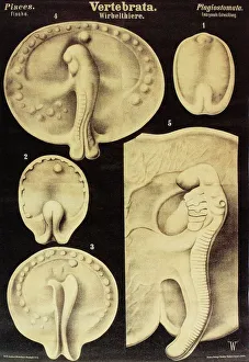
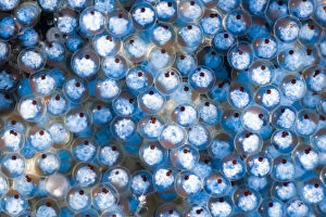
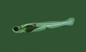
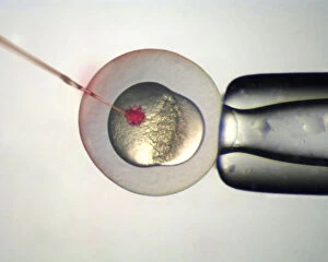
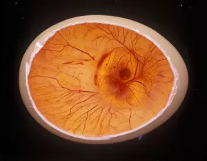
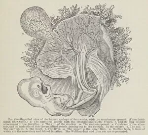
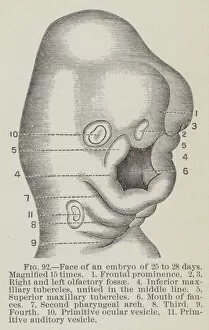
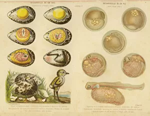
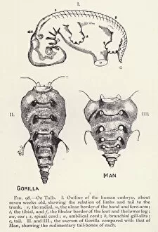
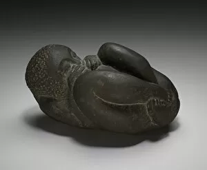
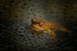
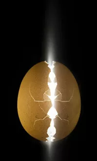
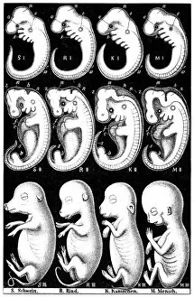
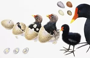
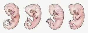
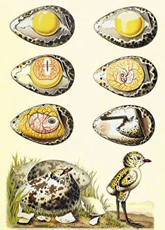
Embryo - a tiny marvel of life, the beginning of a new existence. In the realm of human biology, the term "embryo" refers to a stage in early development known as the human blastocyst. This remarkable phase marks the formation of various cell types that will eventually give rise to all organs and tissues. But humans are not alone in this journey from conception to birth. Comparative embryology sheds light on our shared ancestry with other species. A fascinating chart created by the Department of Comparative and Human Anatomy at the American Museum of Natural History in 1932 showcases this connection. From fish to man, it reveals striking similarities in embryonic development across different organisms. Take, for instance, a chicken chick nestled within its eggshell after twenty days of incubation. At this point, feathers begin to emerge as delicate wings take shape beneath their protective shell. Similarly captivating is an image capturing developing trout eggs; each one holds immense potential for growth and survival. Pictures numbered 11675528 through 11675525 offer glimpses into these diverse embryos' intricate structures and stages of maturation—a testament to nature's incredible diversity and complexity. Meanwhile, another snapshot displays a ten-day-old chicken embryo still enclosed within its egg—an awe-inspiring sight showcasing how life unfolds even before hatching occurs. Zooming closer into human reproduction, we encounter an astonishing photograph revealing embryo development just 24-36 hours after fertilization (Picture No. 12019792). It captures those crucial initial moments when cells rapidly divide and multiply—laying down foundations for future growth. Lastly, let us not forget about our own kind—the miracle that takes place inside every woman's womb: a human fetus cradled within its amniotic sac (Picture No. XXXXXXXX). As it floats weightlessly amidst nourishing fluids, vital organs form gradually while tiny limbs grow stronger day by day—a testament to both resilience and vulnerability.