Premium Framed Print > Science > SEM
Premium Framed Print : Coloured SEM of a section through nerve fibres


![]()
Framed Photos from Science Photo Library
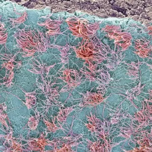
Coloured SEM of a section through nerve fibres
Nerve fibres. Coloured scanning electron micrograph (SEM) of a section through bundles of nerve fibres (red). Each is within a sheath, or perineurium (blue). The nerve fibre bundles (fasciculi) are held together by connective tissue (green). Each fibre consists of a nerve cell axon, the output process of a nerve cell, surrounded by a fatty insulating layer known as a myelin sheath. Myelin sheaths increase the transm- ission speed of the electrical nerve signals. Nerves in the body serve to collect, interpret and relay information. The fibres pass the information on to other parts of the body, such as the muscles and organs. Magnification unknown
Science Photo Library features Science and Medical images including photos and illustrations
Media ID 6421760
© STEVE GSCHMEISSNER/SCIENCE PHOTO LIBRARY
Axon Connective Tissue Fasciculus Fibre Fibres Nerve Nerve Fibre Nerve Fibres Nervous Perineurium System
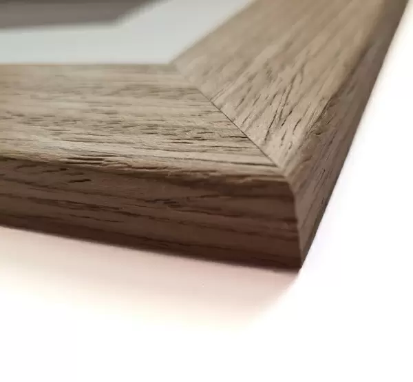
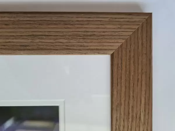
23"x19" (58x48cm) Premium Frame
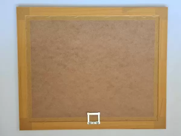
FSC real wood frame with double mounted 16x12 print. Double mounted with white conservation mountboard. Frame moulding comprises stained composite natural wood veneers (Finger Jointed Pine) 39mm wide by 21mm thick. Archival quality Fujifilm CA photo paper mounted onto 1mm card. Overall outside dimensions are 23x19 inches (584x482mm). Rear features Framing tape to cover staples, 50mm Hanger plate, cork bumpers. Glazed with durable thick 2mm Acrylic to provide a virtually unbreakable glass-like finish. Acrylic Glass is far safer, more flexible and much lighter than typical mineral glass. Moreover, its higher translucency makes it a perfect carrier for photo prints. Acrylic allows a little more light to penetrate the surface than conventional glass and absorbs UV rays so that the image and the picture quality doesn't suffer under direct sunlight even after many years. Easily cleaned with a damp cloth. Please note that, to prevent the paper falling through the mount window and to prevent cropping of the original artwork, the visible print may be slightly smaller to allow the paper to be securely attached to the mount without any white edging showing and to match the aspect ratio of the original artwork.
FSC Real Wood Frame and Double Mounted with White Conservation Mountboard - Professionally Made and Ready to Hang
Estimated Image Size (if not cropped) is 27cm x 39.6cm (10.6" x 15.6")
Estimated Product Size is 48.2cm x 58.4cm (19" x 23")
These are individually made so all sizes are approximate
Artwork printed orientated as per the preview above, with portrait (vertical) orientation to match the source image.
FEATURES IN THESE COLLECTIONS
EDITORS COMMENTS
This print showcases the intricate beauty of nerve fibres within the human body. In this coloured scanning electron micrograph (SEM), we are granted a glimpse into the complex network that allows our nervous system to function seamlessly. The image reveals bundles of nerve fibres, depicted in striking red hues, each encased within a protective sheath called perineurium, represented by shades of blue. These fibre bundles, known as fasciculi, are held together by vibrant green connective tissue. At closer inspection, we can observe that every individual fibre consists of a nerve cell axon enveloped by a fatty insulating layer called myelin sheath. This myelin sheath plays a crucial role in increasing the speed at which electrical signals are transmitted along the nerves. Nerves serve as messengers within our bodies, collecting and interpreting information before relaying it to various parts such as muscles and organs. They form an integral part of our sensory perception and motor functions. This mesmerizing photograph not only highlights the incredible complexity and organization found within our nervous system but also serves as a reminder of how interconnected and remarkable our own bodies truly are.
MADE IN THE UK
Safe Shipping with 30 Day Money Back Guarantee
FREE PERSONALISATION*
We are proud to offer a range of customisation features including Personalised Captions, Color Filters and Picture Zoom Tools
SECURE PAYMENTS
We happily accept a wide range of payment options so you can pay for the things you need in the way that is most convenient for you
* Options may vary by product and licensing agreement. Zoomed Pictures can be adjusted in the Basket.