Photographic Print : Skin anatomy

![]()
Photo Prints from Science Photo Library
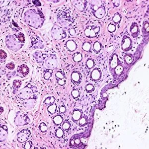
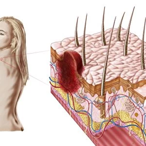
Skin anatomy
Skin anatomy. Historical anatomical artwork of the human skin. The main diagram (lower centre) shows scalp hairs and sebaceous glands (yellow, near surface). The upper diagram shows ridges on skin from the hands (which gives rise to fingerprints). It also shows sweat ducts and pores. Skin blood vessels (upper left) and lymphatic vessels (upper right) are shown, as is the structure of the nails (side view at centre left; top view with the nail removed at centre right). The underside of a nail is at bottom left. The capillaries at bottom right show the structure of papillae (the touch sense organs). Artwork from The Nerves of the Human Body (Ed. Jones Quain, London, 1839)
Science Photo Library features Science and Medical images including photos and illustrations
Media ID 6448465
© SHEILA TERRY/SCIENCE PHOTO LIBRARY
1839 Blood Vessels Book Drawing Duct Ducts Finger Nail Finger Print Forensic Forensics Gland Glands Hair Hairs Hand Jones Quain Lymphatic Nail Nails Papilla Papillae Pore Pores Scalp Sense Skin Sweat Text Book Touch Vessel Finger Nails Section Sectioned
12"x8" (30x20cm) Photo Print
Discover the intricacies of human anatomy with our Media Storehouse Photographic Prints featuring the captivating image "Skin Anatomy" by Science Photo Library. This historical artwork offers a unique glimpse into the complex structure of the skin, with a focus on scalp hairs and sebaceous glands. Bring the wonders of science into your home or office with our high-quality, museum-grade prints. Each print is meticulously produced using archival inks and premium paper to ensure lasting beauty and vibrancy. Order now and ignite your curiosity with this stunning representation of the body's largest organ.
Printed on archival quality paper for unrivalled stable artwork permanence and brilliant colour reproduction with accurate colour rendition and smooth tones. Printed on professional 234gsm Fujifilm Crystal Archive DP II paper. 12x8 for landscape images, 8x12 for portrait images.
Our Photo Prints are in a large range of sizes and are printed on Archival Quality Paper for excellent colour reproduction and longevity. They are ideal for framing (our Framed Prints use these) at a reasonable cost. Alternatives include cheaper Poster Prints and higher quality Fine Art Paper, the choice of which is largely dependant on your budget.
Estimated Image Size (if not cropped) is 19.1cm x 30.4cm (7.5" x 12")
Estimated Product Size is 20.3cm x 30.5cm (8" x 12")
These are individually made so all sizes are approximate
Artwork printed orientated as per the preview above, with portrait (vertical) orientation to match the source image.
EDITORS COMMENTS
This print showcases a remarkable piece of historical anatomical artwork depicting the intricate details of human skin. Dating back to 1839, this illustration from "The Nerves of the Human Body" by Jones Quain offers a fascinating glimpse into the understanding of skin anatomy during that era. In the main diagram at the lower center, one can observe scalp hairs and sebaceous glands near the surface, depicted in a vibrant yellow hue. The upper diagram highlights ridges on the hands which give rise to fingerprints, along with sweat ducts and pores. Additionally, it provides an insight into skin blood vessels on the upper left and lymphatic vessels on the upper right. The structure of nails is also illustrated here, showcasing both side view and top view perspectives. Delving deeper into this masterpiece reveals intriguing details such as papillae - touch sense organs - showcased through capillaries at bottom right. Furthermore, forensic enthusiasts will appreciate how this artwork captures essential elements for identification purposes like finger prints and nail structures. As we admire this stunning artistic representation steeped in medical history, we are reminded of its significance as a valuable resource for scientific education and research. This image serves as a testament to humanity's continuous exploration of our own bodies throughout time – an enduring pursuit that has shaped our understanding of medicine and anatomy today.
MADE IN THE UK
Safe Shipping with 30 Day Money Back Guarantee
FREE PERSONALISATION*
We are proud to offer a range of customisation features including Personalised Captions, Color Filters and Picture Zoom Tools
SECURE PAYMENTS
We happily accept a wide range of payment options so you can pay for the things you need in the way that is most convenient for you
* Options may vary by product and licensing agreement. Zoomed Pictures can be adjusted in the Basket.