Fraud Blocker Skip to main content

Pore Collection

"Pore: A Window into the Intricacies of Nature's Design" The term "pore" may evoke thoughts of skincare or beauty products, but in the realm of science and nature

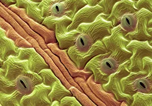
Background imagePore Collection: Leaf pores, SEM

Leaf pores, SEM
Leaf pores. Coloured scanning electron micrograph (SEM) of stomata (holes) on the surface of a leaf. These structures perform a similar function to the pores in human skin

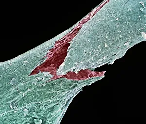
Background imagePore Collection: Osteoporotic bone, SEM

Osteoporotic bone, SEM
Osteoporotic bone. Coloured scanning electron micrograph (SEM) of fractured bone tissue from a patient suffering from osteoporosis (brittle bone disease)

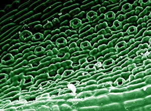
Background imagePore Collection: Picture No. 11675585

Picture No. 11675585
Scanning Electron micrograph (SEM)showing stomata on a Yew Leaf. Date:

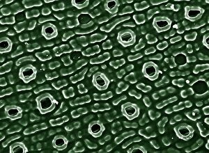
Background imagePore Collection: English oak leaf pores, SEM

English oak leaf pores, SEM
English oak leaf pores. Coloured scanning electron micrograph (SEM) of stomata (round) on the underside of a leaf from an English oak (Quercus robur) tree

Background imagePore Collection: Picture No. 11675628

Picture No. 11675628
Scanning Electron Micrograph (SEM): Stomata of Yew Leaf. Date:

Background imagePore Collection: French lavender leaf pore, SEM

French lavender leaf pore, SEM
French lavender leaf pore. Coloured scanning electron micrograph (SEM) of an open stoma (centre, black). Stomata are pores that open and close in order to regulate gas exchange in a plant

Background imagePore Collection: Sunset at Giau Pass, Colle Santa Lucia, Belluno district, Veneto, Italy

Sunset at Giau Pass, Colle Santa Lucia, Belluno district, Veneto, Italy

Background imagePore Collection: Mount Pore and Mount Civetta at sunset, Colle Santa Lucia, Belluno district, Veneto, Italy

Mount Pore and Mount Civetta at sunset, Colle Santa Lucia, Belluno district, Veneto, Italy

Background imagePore Collection: Mount Civetta and Mount Pore at sunset, Alleghe, Belluno district, Veneto, Italy

Mount Civetta and Mount Pore at sunset, Alleghe, Belluno district, Veneto, Italy

Background imagePore Collection: Grey Reef Shark

Grey Reef Shark
A close look at the sensory organs known as ampullae of Lorenzini, on a Grey reef shark (Carcharhinus amblyrhynchos. These dark pores around their snout and face pick up electrical fields)

Background imagePore Collection: Close Up of the Seed Head of a Dandelion

Close Up of the Seed Head of a Dandelion
Dandelion is a large genus of flowering plants in the family Asteraceae and are native to Eurasia and North America. They have very small flowers collected together into a composite flower head

Background imagePore Collection: Digital illustration of Spiny Anteater (Echidna) puggle sucking from pores on milk patch in pouch

Digital illustration of Spiny Anteater (Echidna) puggle sucking from pores on milk patch in pouch

Background imagePore Collection: Cell membrane ion channels, artwork C016 / 7689

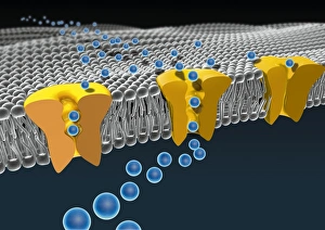
Cell membrane ion channels, artwork C016 / 7689
Cell membrane ion channels. Computer artwork of a section through the membrane of an animal cell, showing transmembrane ion channel proteins (yellow)

Background imagePore Collection: Chives leaf stoma, SEM C016 / 8060

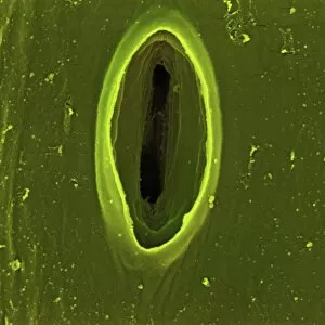
Chives leaf stoma, SEM C016 / 8060
Chives leaf stoma. Coloured scanning electron micrograph (SEM) of a stoma (oval, centre) on the surface of a leaf from a chives (Allium schoenoprasum) plant

Background imagePore Collection: Coltsfoot leaf stomata, SEM C016 / 8052

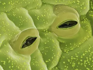
Coltsfoot leaf stomata, SEM C016 / 8052
Coltsfoot leaf stomata. Coloured scanning electron micrograph (SEM) of two stomata (round) on the surface of a leaf from a coltsfoot (Tussilago farfara) plant

Background imagePore Collection: Potato Leaf Stomata (SEM)

Potato Leaf Stomata (SEM)
Potato Stomata. Scanning electron micrographs (SEM) of open stomata on a potato leaf (Solanum tuberosum). Stomata are pores that open and close in order to regulate gas exchange in a plant

Background imagePore Collection: Human skin seen under a microscope, showing hair shaft and sweat pores

Human skin seen under a microscope, showing hair shaft and sweat pores

Background imagePore Collection: Picture No. 11675633

Picture No. 11675633
Light Micrograph (LM): A transverse section of a stem of Clover (Trifolium sp.) with an unidentified parasite attached. Date:

Background imagePore Collection: Microscopic view of plant tissues

Microscopic view of plant tissues

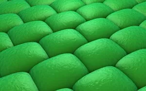
Background imagePore Collection: Conceptual image of stomata

Conceptual image of stomata

Background imagePore Collection: Red-knobbed star (Fromia elegans) on Pore Coral, Indian Ocean, Maldives

Red-knobbed star (Fromia elegans) on Pore Coral, Indian Ocean, Maldives

Background imagePore Collection: Brown-rot (Postia placenta) close-up detail of pores, Clumber Park, Nottinghamshire, England

Brown-rot (Postia placenta) close-up detail of pores, Clumber Park, Nottinghamshire, England, October

Background imagePore Collection: Pseudocrinites magnificus, cystoid fossilized in Wenlock limestone

Pseudocrinites magnificus, cystoid fossilized in Wenlock limestone, featuring rhombic respiratory pore

Background imagePore Collection: Porous brown surface

Porous brown surface

Background imagePore Collection: Fruit fly spiracle, SEM

Fruit fly spiracle, SEM
Fruit fly (Drosophila melanogaster) spiracle. Coloured scanning electron micrograph (SEM) of the spiracle (air pore) of a male fruit fly

Background imagePore Collection: Radiolarian skeleton, artwork F005 / 0194

Radiolarian skeleton, artwork F005 / 0194
Radiolarian skeleton. Computer artwork of the skeleton of a radiolarian. Radiolaria are single-celled protozoans that are found in marine plankton

Background imagePore Collection: Radiolarian skeleton, artwork F005 / 0190

Radiolarian skeleton, artwork F005 / 0190
Radiolarian skeleton. Computer artwork of the skeleton of a radiolarian. Radiolaria are single-celled protozoans that are found in marine plankton

Background imagePore Collection: Human skin anatomy, artwork

Human skin anatomy, artwork
Human skin anatomy, computer artwork

Background imagePore Collection: Anthrax protective antigen molecule C014 / 0886

Anthrax protective antigen molecule C014 / 0886
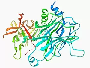
Anthrax protective antigen molecule. Computer model showing the structure of a molecule of protective antigen (PA) produced by anthrax (Bacillus anthracis) bacteria

Background imagePore Collection: Anthrax protective antigen molecule C014 / 0865

Anthrax protective antigen molecule C014 / 0865
Anthrax protective antigen molecule. Computer model showing the structure of a molecule of protective antigen (PA) produced by anthrax (Bacillus anthracis) bacteria

Background imagePore Collection: Nucleus and endoplasmic reticulum F006 / 9196

Nucleus and endoplasmic reticulum F006 / 9196
Computer artwork showing part of a human or eukaryotic cell. In the middle the nucleus which has a membrane with nuclear pores. Inside the nucleus is the DNA

Background imagePore Collection: Voltage-gated potassium channel F006 / 9642

Voltage-gated potassium channel F006 / 9642
Voltage-gated potassium channel. Molecular model of a voltage-gated potassium (Kv) ion channel. Ion channels are membrane-spanning proteins that form pores in cell membranes

Background imagePore Collection: Voltage-gated potassium channel F006 / 9562

Voltage-gated potassium channel F006 / 9562
Voltage-gated potassium (Kv) ion channel, molecular model. Ion channels are membrane-spanning proteins that form pores in cell membranes

Background imagePore Collection: Voltage-gated potassium channel F006 / 9391

Voltage-gated potassium channel F006 / 9391
Voltage-gated potassium channel. Molecular model of a voltage-gated potassium (Kv) ion channel complexed with the antigen-binding fragment (Fab) of a monoclonal antibody

Background imagePore Collection: Voltage-gated potassium channel F006 / 9324

Voltage-gated potassium channel F006 / 9324
Voltage-gated potassium channel. Molecular model of a voltage-gated potassium (Kv) ion channel. Ion channels are membrane-spanning proteins that form pores in cell membranes

Background imagePore Collection: Anthrax protective antigen molecule F006 / 9229

Anthrax protective antigen molecule F006 / 9229
Anthrax protective antigen molecule. Computer model showing the structure of a molecule of protective antigen (PA) produced by anthrax (Bacillus anthracis) bacteria

Background imagePore Collection: Anthrax protective antigen molecule F006 / 9225

Anthrax protective antigen molecule F006 / 9225
Anthrax protective antigen molecule. Computer model showing the structure of a molecule of protective antigen (PA) produced by anthrax (Bacillus anthracis) bacteria

Background imagePore Collection: Nucleus and endoplasmic reticulum F006 / 9201

Nucleus and endoplasmic reticulum F006 / 9201
Computer artwork showing part of a human or eukaryotic cell. In the middle the nucleus which has a membrane with nuclear pores. Inside the nucleus is the DNA

Background imagePore Collection: Tobacco leaf, SEM C016 / 9465

Tobacco leaf, SEM C016 / 9465
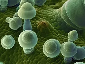
Tobacco leaf. Coloured scanning electron micrograph (SEM) of the underside of a winged tobacco (Nicotiana alata) leaf, showing trichomes (long) and a stomata (small, oval)

Background imagePore Collection: Squash leaf, SEM C016 / 9463

Squash leaf, SEM C016 / 9463
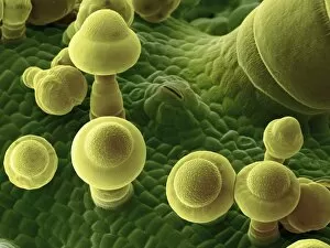
Squash leaf. Coloured scanning electron micrograph (SEM) of the lower surface of a squash (Curcubita maxima) leaf, showing trichomes (mushroom-shaped) and a stoma (slit, upper centre)

Background imagePore Collection: Thale cress leaf stoma, SEM C016 / 9460

Thale cress leaf stoma, SEM C016 / 9460
Thale cress leaf stoma. Coloured scanning electron micrograph (SEM) of a stoma (round, centre) on the surface of a leaf from a thale cress (Arabidopsis thaliana) plant

Background imagePore Collection: Thale cress leaf stoma, SEM C016 / 9459

Thale cress leaf stoma, SEM C016 / 9459
Thale cress leaf stoma. Coloured scanning electron micrograph (SEM) of a stoma (round, centre) on the surface of a leaf from a thale cress (Arabidopsis thaliana) plant

Background imagePore Collection: Skin structure, artwork C016 / 7541

Skin structure, artwork C016 / 7541
Skin structure. Computer artwork showing the different layers and structure of human skin. The upper layer (centre) is the epidermis, with the dermis below

Background imagePore Collection: Squash leaf, SEM C016 / 9464

Squash leaf, SEM C016 / 9464
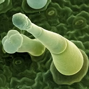
Squash leaf. Coloured scanning electron micrograph (SEM) of the underside of a squash (Curcubita maxima) leaf, showing trichomes (mushroom-shaped) and a stoma (slit, upper centre)

Background imagePore Collection: Ebola matrix protein molecule

Ebola matrix protein molecule
Ebola matrix protein. Molecular model of the Ebola virus matrix protein VP40 (green) bound to RNA (ribonucleic acid, red)

Background imagePore Collection: Anthrax toxin prepore, molecular model

Anthrax toxin prepore, molecular model
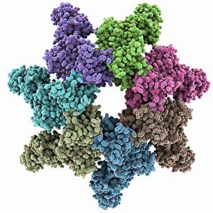
Anthrax toxin prepore. Molecular model of protective antigen (PA) from anthrax (Bacillus anthracis) bacteria complexed with an anthrax toxin receptor, forming a prepore (hole, centre)

Background imagePore Collection: Plant stoma, ESEM

Plant stoma, ESEM
Plant stoma, coloured environmental scanning electron micrograph (ESEM). A stoma is a pore that regulates the exchange of gases and water vapour into and out of the plant


All Professionally Made to Order for Quick Shipping

"Pore: A Window into the Intricacies of Nature's Design" The term "pore" may evoke thoughts of skincare or beauty products, but in the realm of science and nature, it holds a much deeper significance. From osteoporotic bone to leaf pores, these microscopic openings offer us a glimpse into the intricate workings of our world. In the fascinating world of SEM (scanning electron microscopy), we uncover hidden wonders that are otherwise invisible to the naked eye. Picture No. 11675585 reveals osteoporotic bone under intense magnification, showcasing its porous structure and shedding light on its vulnerability. Moving from bones to leaves, Picture No. 11675628 captures English oak leaf pores in stunning detail. These tiny openings play a crucial role in regulating gas exchange within plants, allowing them to breathe and thrive. Venturing further into nature's diversity, we encounter French lavender leaf pore through SEM imagery. The delicate intricacy displayed by this pore reminds us of the remarkable design found even at such minuscule scales. But our journey doesn't end there; it extends beyond microscopic realms to breathtaking landscapes captured at sunset in Giau Pass and Colle Santa Lucia, Belluno district, Veneto, Italy. Witnessing Mount Pore and Mount Civetta bathed in golden hues offers a momentary escape into serene beauty—a reminder of nature's grandeur that surrounds us. As we delve deeper into Alleghe's Belluno district in Veneto, Italy—where Mount Civetta stands tall alongside Mount Pore—we witness another awe-inspiring sunset scene. The interplay between light and shadow paints an ethereal canvas against which these majestic peaks stand as silent witnesses to time passing by. And finally, amidst all these natural wonders lies an unexpected guest—the Grey Reef Shark—an apex predator lurking beneath ocean waves with its own set of unique adaptations for survival. In essence, "pore" encapsulates not just a simple opening but an entire world of discovery.