
Pore Collection (page 3)
"Pore: A Window into the Intricacies of Nature's Design" The term "pore" may evoke thoughts of skincare or beauty products, but in the realm of science and nature


All Professionally Made to Order for Quick Shipping
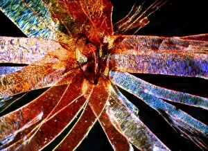
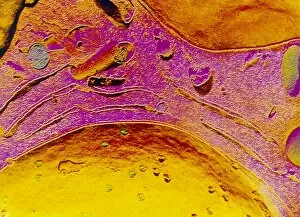
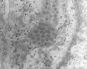
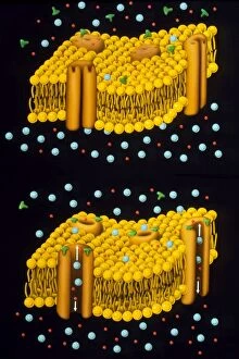
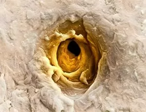
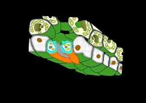
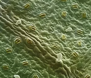
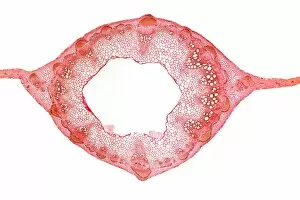
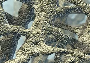
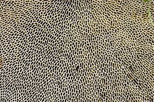
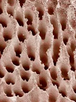
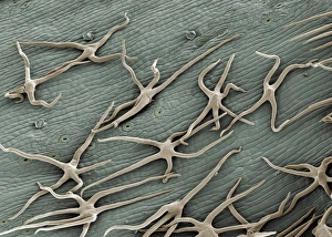
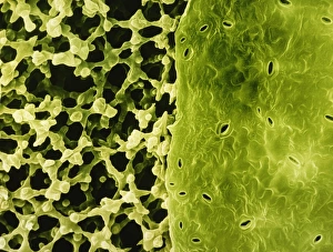
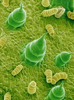
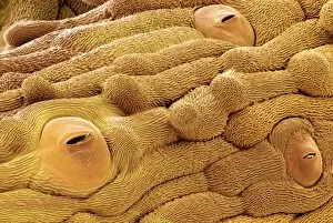
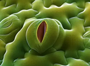
"Pore: A Window into the Intricacies of Nature's Design" The term "pore" may evoke thoughts of skincare or beauty products, but in the realm of science and nature, it holds a much deeper significance. From osteoporotic bone to leaf pores, these microscopic openings offer us a glimpse into the intricate workings of our world. In the fascinating world of SEM (scanning electron microscopy), we uncover hidden wonders that are otherwise invisible to the naked eye. Picture No. 11675585 reveals osteoporotic bone under intense magnification, showcasing its porous structure and shedding light on its vulnerability. Moving from bones to leaves, Picture No. 11675628 captures English oak leaf pores in stunning detail. These tiny openings play a crucial role in regulating gas exchange within plants, allowing them to breathe and thrive. Venturing further into nature's diversity, we encounter French lavender leaf pore through SEM imagery. The delicate intricacy displayed by this pore reminds us of the remarkable design found even at such minuscule scales. But our journey doesn't end there; it extends beyond microscopic realms to breathtaking landscapes captured at sunset in Giau Pass and Colle Santa Lucia, Belluno district, Veneto, Italy. Witnessing Mount Pore and Mount Civetta bathed in golden hues offers a momentary escape into serene beauty—a reminder of nature's grandeur that surrounds us. As we delve deeper into Alleghe's Belluno district in Veneto, Italy—where Mount Civetta stands tall alongside Mount Pore—we witness another awe-inspiring sunset scene. The interplay between light and shadow paints an ethereal canvas against which these majestic peaks stand as silent witnesses to time passing by. And finally, amidst all these natural wonders lies an unexpected guest—the Grey Reef Shark—an apex predator lurking beneath ocean waves with its own set of unique adaptations for survival. In essence, "pore" encapsulates not just a simple opening but an entire world of discovery.