Home > Science > SEM
Sunflower leaf, SEM
![]()

Wall Art and Photo Gifts from Science Photo Library
Sunflower leaf, SEM
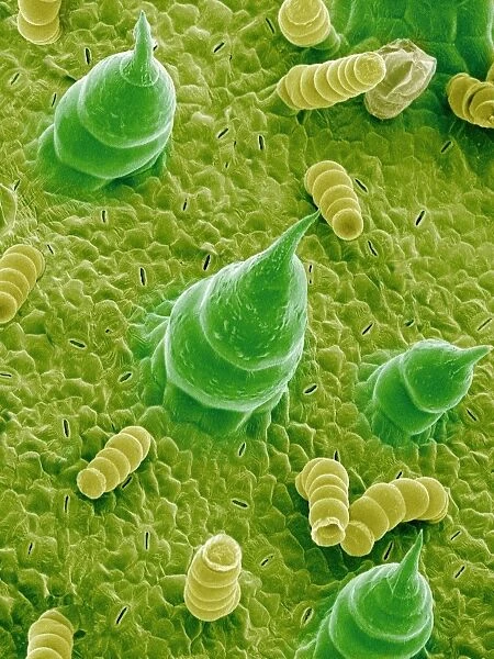
Sunflower leaf. Coloured scanning electron micrograph (SEM) of the underside of a sunflower leaf (Helianthus annuus). The green and yellow structures are trichomes, which protect the plant against predators and reduce water loss through evaporation. Also seen are pores (slits), called stomata. These are the site of gaseous exchange, or respiration. Carbon dioxide enters the plant for use in photosynthesis, and waste oxygen exits the plants tissues at the stomata. Water vapour also evaporates through the stomata. Magnification: x430 when printed at 10 centimetres tall
Science Photo Library features Science and Medical images including photos and illustrations
Media ID 6289173
© SUSUMU NISHINAGA/SCIENCE PHOTO LIBRARY
Gaseous Exchange Hair Hairs Helianthus Annuus Pore Pores Protection Protective Respiration Stoma Stomata Sun Flower Trichome Trichomes Under Side Water Loss False Coloured
EDITORS COMMENTS
This print showcases the intricate beauty of a sunflower leaf like never before. Taken using a scanning electron microscope (SEM), the image reveals the vibrant colors and fascinating structures that make up this botanical wonder. The green and yellow formations are known as trichomes, which play a crucial role in safeguarding the plant from predators while minimizing water loss through evaporation. Upon closer inspection, one can also spot tiny slits called stomata scattered across the leaf's surface. These pores serve as gateways for gaseous exchange or respiration within the plant. As carbon dioxide enters through these openings, it fuels photosynthesis - an essential process for plants to produce energy. Simultaneously, waste oxygen is expelled from the plant's tissues via these stomata. The SEM magnification of x430 used to capture this image allows us to appreciate every minute detail when printed at 10 centimeters tall. It provides viewers with an extraordinary glimpse into nature's ingenious design and reminds us of its complexity on a microscopic level. This photograph not only celebrates botany but also highlights how plants have evolved various mechanisms, such as trichomes and stomata, to adapt and thrive in their environments. It serves as a testament to both the resilience and delicate balance found within our natural world.
MADE IN THE UK
Safe Shipping with 30 Day Money Back Guarantee
FREE PERSONALISATION*
We are proud to offer a range of customisation features including Personalised Captions, Color Filters and Picture Zoom Tools
SECURE PAYMENTS
We happily accept a wide range of payment options so you can pay for the things you need in the way that is most convenient for you
* Options may vary by product and licensing agreement. Zoomed Pictures can be adjusted in the Basket.


