Fraud Blocker Skip to main content

Papillae Collection

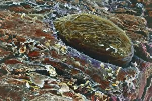
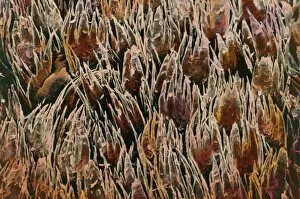
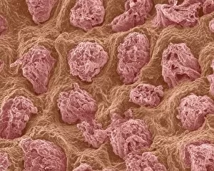
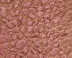
"Papillae: Unveiling the Intricate World of Texture and Taste" Discover the fascinating realm of papillae, as we delve into their diverse forms and functions

Background imagePapillae Collection: Cat tongue surface, SEM

Cat tongue surface, SEM
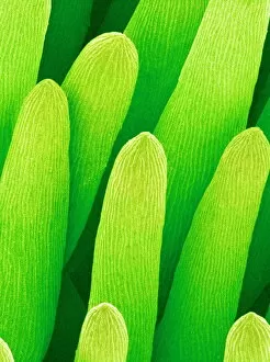
Cats tongue. Coloured scanning electron micrograph (SEM) of the surface of a cats (family Felidae) tongue. The tongue is covered in many backward facing papillae (projections)

Background imagePapillae Collection: Forget-me-not flower, SEM

Forget-me-not flower, SEM
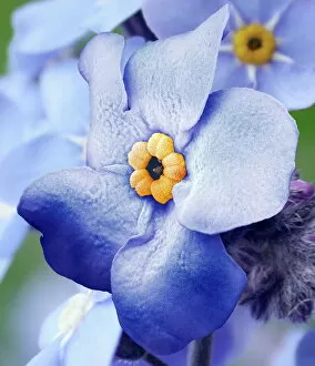
Forget-me-not flower. Coloured scanning electron micrograph (SEM) of a forget-me-not (Myosotis arvensis) flower. The corolla consists of five petals (blue)

Background imagePapillae Collection: Digital cross section illustration of taste buds and filiform papilae on tongue

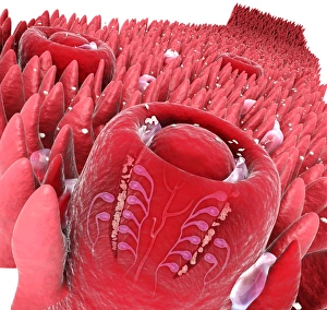
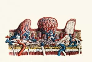
Digital cross section illustration of taste buds and filiform papilae on tongue

Background imagePapillae Collection: Forget-me-not flower, SEM

Forget-me-not flower, SEM
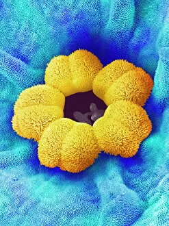
Forget-me-not flower. Coloured scanning electron micrograph (SEM) of a forget-me-not (Myosotis arvensis) flower. The petals (blue) are covered by numerous projections called papillae

Background imagePapillae Collection: Yarrow flower petal, SEM

Yarrow flower petal, SEM
Yarrow flower petal. Coloured scanning electron micrograph (SEM) of a petal from the flower of a Japanese type of yarrow (Achillea alpina)

Background imagePapillae Collection: Zinnia flower petal, SEM

Zinnia flower petal, SEM
Zinnia flower petal. Coloured scanning electron micrograph (SEM) of a Zinnia sp. petal. The projections on the petal help to prevent water loss

Background imagePapillae Collection: Cow tongue under the microscope

Cow tongue under the microscope: veal tongue showing different papillae 1, foliate papilla 2, filiform papillae 3, and fungiform papillae 4

Background imagePapillae Collection: Picture No. 11675366

Picture No. 11675366
Flatworm. Date:

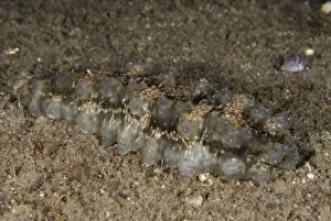
Background imagePapillae Collection: Picture No. 11675345

Picture No. 11675345
Dragonfish Sea Cucumber with extended papillae on black sand. Date:

Background imagePapillae Collection: Picture No. 11072772

Picture No. 11072772
White-V Octopus camouflaged on black sand TK1 dive site, Lembeh Straits, Sulawesi, Indonesia. White-V Octopus

Background imagePapillae Collection: Picture No. 11072723

Picture No. 11072723
Pair of Bigfin Reef Squid during night dive Batu Angus dive site, Lembeh Straits, Sulawesi, Indonesia. Pair of Bigfin Reef Squid

Background imagePapillae Collection: Picture No. 11072719

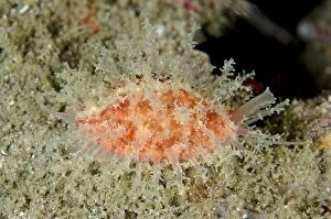
Picture No. 11072719
Cowrie with extended papillae Tanjung Slope dive site, Lembeh Straits, Sulawesi, Indonesia. Cowrie

Background imagePapillae Collection: Picture No. 11072907

Picture No. 11072907
Bigfin Reef Squid on nght dive TK1 dive site, Lembeh Straits, Sulawesi, Indonesia. Bigfin Reef Squid

Background imagePapillae Collection: Picture No. 11072880

Picture No. 11072880
Veined Octopus with leaf on black sand on night dive Jahir dive site, Lembeh Straits, Sulawesi, Indonesia. Veined Octopus

Background imagePapillae Collection: Picture No. 10877011

Picture No. 10877011
Scanning Electron Micrograph (SEM): Frog tongue - taste receptors papillae Date:

Background imagePapillae Collection: Tongue, Keratin that covers the apex of the papillae under microscope

Tongue, Keratin that covers the apex of the papillae under microscope
Medicine - Human Histology - Tongue - Keratin that covers the apex of the papillae under microscope

Background imagePapillae Collection: Tongue, Vallate papillae under microscope

Tongue, Vallate papillae under microscope
Medicine - Human Histology - Tongue - Vallate papillae under microscope

Background imagePapillae Collection: Tongue, Fungiform papillae under microscope

Tongue, Fungiform papillae under microscope
Medicine - Human Histology - Tongue - Fungiform papillae under microscope

Background imagePapillae Collection: Tongue, Filiform papillae under microscope

Tongue, Filiform papillae under microscope
Medicine - Human Histology - Tongue - Filiform papillae under microscope

Background imagePapillae Collection: Kidney anatomy, artwork C016 / 8945

Kidney anatomy, artwork C016 / 8945
Kidney anatomy, artwork. The kidneys filter waste from the blood and excrete it as urine. The kidney consists of an outer cortex and an inner medulla

Background imagePapillae Collection: Pea flower pollination, SEM C016 / 0067

Pea flower pollination, SEM C016 / 0067
Pea flower pollination. Coloured scanning electron micrograph (SEM) of pollen grains (ovals) on the stigma of a garden pea flower (Pisum sativum)

Background imagePapillae Collection: Tongue, light micrograph C015 / 7105

Tongue, light micrograph C015 / 7105
Tongue. Light micrograph of a section through healthy tongue tissue. The surface of the tongue (across top) consists of stratified squamous epithelium (dark purple) with a rough keratinised surface

Background imagePapillae Collection: Pea flower pollination, SEM C016 / 0065

Pea flower pollination, SEM C016 / 0065
Pea flower pollination. Coloured scanning electron micrograph (SEM) of pollen grains (ovals) on the stigma of a garden pea flower (Pisum sativum)

Background imagePapillae Collection: Pea flower pollination, SEM C016 / 0066

Pea flower pollination, SEM C016 / 0066
Pea flower pollination. Coloured scanning electron micrograph (SEM) of pollen grains (ovals) on the stigma of a garden pea flower (Pisum sativum)

Background imagePapillae Collection: Human tongue, SEM C014 / 4855

Human tongue, SEM C014 / 4855
Human tongue. Coloured scanning electron micrograph (SEM) showing the surface structure of a human tongue. The taste-buds (papillae)

Background imagePapillae Collection: Human tongue, SEM C014 / 4854

Human tongue, SEM C014 / 4854
Human tongue. Coloured scanning electron micrograph (SEM) showing the surface structure of a human tongue. The taste-buds (papillae)

Background imagePapillae Collection: Kidney anatomy, artwork C016 / 8663

Kidney anatomy, artwork C016 / 8663
Kidney anatomy, artwork. The kidneys filter waste from the blood and excrete it as urine. The kidney consists of an outer cortex and an inner medulla

Background imagePapillae Collection: Kidney anatomy, artwork C016 / 8661

Kidney anatomy, artwork C016 / 8661
Kidney anatomy, artwork. The kidneys filter waste from the blood and excrete it as urine. The kidney consists of an outer cortex and an inner medulla

Background imagePapillae Collection: Kidney anatomy, artwork C016 / 8662

Kidney anatomy, artwork C016 / 8662
Kidney anatomy, artwork. The kidneys filter waste from the blood and excrete it as urine. The kidney consists of an outer cortex and an inner medulla

Background imagePapillae Collection: Kidney anatomy, artwork C016 / 8660

Kidney anatomy, artwork C016 / 8660
Kidney anatomy, artwork. The kidneys filter waste from the blood and excrete it as urine. The kidney consists of an outer cortex and an inner medulla

Background imagePapillae Collection: Horseradish petal, SEM C016 / 8062

Horseradish petal, SEM C016 / 8062
Horseradish petal. Coloured scanning electron micrograph (SEM) of a petal from a horseradish (Armoracia rusticana) flower, showing the structure of the papillae that make up its surface

Background imagePapillae Collection: Petal papillae, SEM

Petal papillae, SEM
Petal papillae. Coloured scanning electron micrograph (SEM) of papillae on the surface of a cultivated primrose (Primula obconica) petal. Papillae are projections from epidermal cells

Background imagePapillae Collection: Petal papillae on a Snapdragon flower

Petal papillae on a Snapdragon flower
Petal papillae. Coloured scanning electron micrograph showing papillae on the petal of a Snapdragon flower, Antirrhinum majus. Papillae are projections from epidermal cells

Background imagePapillae Collection: Hedgehog tongue, SEM

Hedgehog tongue, SEM
Hedgehog tongue. Coloured scanning electron micrograph (SEM) of filiform papillae on the surface of a hedgehogs tongue. These small projections are used for grooming

Background imagePapillae Collection: Mole nose, SEM

Mole nose, SEM
Mole nose. Coloured scanning electron micrograph (SEM) of the snout (pink) of a mole, showing specialised bulbous papillae (projections) known as Eimers organs

Background imagePapillae Collection: Papillae on the tongue of a bat

Papillae on the tongue of a bat
Papillae on the tongue of a pipistrelle bat. Coloured scanning electron micrograph (SEM) of the tongue of a pipistrelle bat (Pipistrellus pipistrellus)

Background imagePapillae Collection: Kites toe papillae, SEM

Kites toe papillae, SEM
Kites toe papillae. Coloured scanning electron micrograph (SEM) of papillae from the base of a kites (family Accipitridae) toe

Background imagePapillae Collection: Pipistrelle bat tongue, SEM

Pipistrelle bat tongue, SEM
Pipistrelle bat tongue. Coloured scanning electron micrograph (SEM) of the surface of a pipistrelle bats (Pipistrellus pipistrellus) tongue showing three types of papillae (projections)

Background imagePapillae Collection: Breathing tube on a fruit flys pupa, SEM

Breathing tube on a fruit flys pupa, SEM
Breathing tube on a fruit flys pupa, coloured scanning electron micrograph (SEM). This structure is called a spiracle. This fruit fly is Drosophila melanogaster (wild type Oregon R)

Background imagePapillae Collection: Tongue papillae, SEM

Tongue papillae, SEM
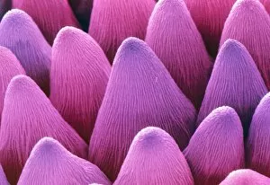
Tongue papillae. Coloured scanning electron micrograph (SEM) of filiform papillae on the surface of the tongue. Some types of papillae, or small projections, house tastebuds

Background imagePapillae Collection: Skin anatomy

Skin anatomy. Historical anatomical artwork of the human skin. The main diagram (lower centre) shows scalp hairs and sebaceous glands (yellow, near surface)

Background imagePapillae Collection: Papillae on the tongue

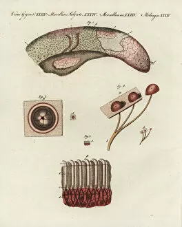
Papillae on the tongue
Papillae on tongue, coloured historical artwork. On the tongue most tastebuds, the structures that provide information about the taste of food, sit on small platforms called papillae

Background imagePapillae Collection: Tongue anatomy

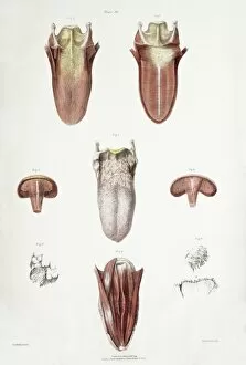
Tongue anatomy. Historical anatomical artwork of a human tongue. The main diagram (centre) shows the upper surface of the tongue

Background imagePapillae Collection: Rabbit tongues surface, SEM

Rabbit tongues surface, SEM
Rabbit tongues surface, coloured scanning electron micrograph (SEM). The tongue is covered in thin projections called filiform papillae. These sense touch

Background imagePapillae Collection: Tongue papillae, light micrograph

Tongue papillae, light micrograph
Tongue papillae. Coloured light micrograph of a section through the tongue, showing its filiform papillae (red). They project from the tongues upper surface, giving it its rough texture

Background imagePapillae Collection: Taste bud, light micrograph

Taste bud, light micrograph
Taste bud. Coloured light micrograph of a section through the tongue, showing a taste bud. This taste bud is seen as a round purple mass slightly to the right of the vertical white line

Background imagePapillae Collection: Taste bud, SEM

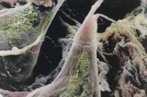
Taste bud, SEM
Taste bud. Coloured scanning electron micrograph (SEM) of a taste bud (centre) on a tongue. This is the centre of the round tongue structure called a fungiform papilla

Background imagePapillae Collection: False-colour SEM of dorsal surface of tongue

False-colour SEM of dorsal surface of tongue
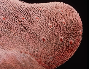
Tongue surface. False-colour scanning electron micrograph (SEM) of the dorsal surface of the tongue. It is covered by two types of projections known as filiform and fungiform papillae


All Professionally Made to Order for Quick Shipping

"Papillae: Unveiling the Intricate World of Texture and Taste" Discover the fascinating realm of papillae, as we delve into their diverse forms and functions. These tiny structures, resembling cat tongue surfaces under a scanning electron microscope (SEM), are not only found in feline friends but also play a crucial role in our own gustatory experiences. Intriguingly, SEM images reveal that papillae extend beyond the realms of animals. Take a closer look at the delicate petals of forget-me-not flowers or yarrow flowers, magnified to showcase their intricate textures reminiscent of filiform papilae on our tongues. The digital cross-section illustration further highlights how taste buds intertwine with these unique projections, forming an essential part of our sensory perception. Zooming back into nature's palette, SEM captures mesmerizing details within zinnia flower petals and pansy flower petals. These close-ups unveil an astonishing array of patterns and textures akin to the microscopic world residing on our tongues. But it doesn't stop there. Let's shift gears from flora to fauna as we explore cow tongue under the microscope. Picture No. 11675366 showcases this intriguing organ up close, revealing its own set of distinctive papillae formations that aid bovines in grazing and consuming vegetation effectively. As we immerse ourselves in these captivating visuals (Picture No. 11675345), let us appreciate how something so seemingly insignificant can hold such profound significance for both humans and animals alike. Papillae serve as gatekeepers to flavor sensations; they allow us to savor every delectable bite while ensuring cows graze efficiently on lush pastures. So next time you enjoy your favorite dish or marvel at nature's floral wonders, remember that beneath it all lies a hidden world shaped by intricate papillae – guardians guiding us through tactile exploration and enhancing our connection with food and beauty (Picture No. 11091797, Picture No. 11072772).