
Lymphatic Collection
The lymphatic system is a complex network of vessels and organs that play a crucial role in our body's immune defense


All Professionally Made to Order for Quick Shipping
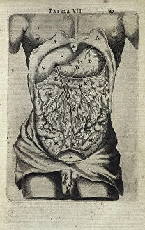
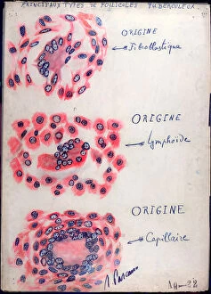
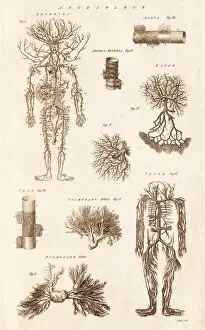
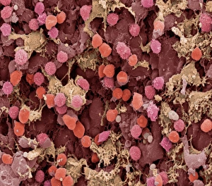
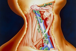
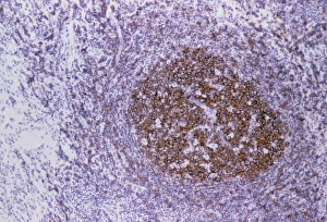
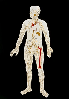
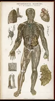
The lymphatic system is a complex network of vessels and organs that play a crucial role in our body's immune defense. Dendritic cells, known as the sentinels of the immune system, are key players in this intricate system. Their ability to capture and present antigens to other immune cells helps initiate an effective immune response. In principle tuberculosis follicles, depicted beautifully through pen and ink on paper artwork, we can see how these tiny structures form within the lymph nodes during infection. This illustration serves as a reminder of the importance of understanding the lymphatic system's role in combating diseases. Moving from the arm to the thorax and abdomen, we witness how this extensive network extends throughout our entire body. The lymphatic vessels transport excess fluid, called lymph, back into circulation while also filtering out harmful substances along with it. Artwork showcasing the lymphatic system in different parts of our body highlights its significance even further. From illustrations depicting its presence in legs and feet to those focusing on arm anatomy, each piece captures its complexity with precision (such as artwork C013 / 4583 or Picture No. 11675487). An illustration portraying both conducting systems and various lymph nodes provides us with a comprehensive view of how this intricate web connects different parts of our body together for optimal functioning. Through captivating artwork dedicated solely to representing the beauty and intricacy of this vital bodily system (like "Lymphatic System" artworks), we gain an appreciation for its essential role beyond what meets the eye. Understanding and appreciating our lymphatic system not only enhances our knowledge but also reminds us to take care of ourselves by maintaining good health practices that support its proper function.