Mounted Print : Skin inflammation, light micrograph


![]()
Mounted Prints from Science Photo Library
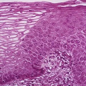
Skin inflammation, light micrograph
Skin inflammation. Light micrograph of a section through skin showing inflamed capillaries (lower left and upper right) caused by capillaritis, also known as pigmented purpurea. The capillary walls become inflamed and blood cells leak out into the surrounding tissues. This leads to reddish brown patches on the surface of the skin. Capillaritis may be caused by an allergic reaction to medication, food additives or a viral infection. There is no known cure except avoidance of possible allergens
Science Photo Library features Science and Medical images including photos and illustrations
Media ID 6413602
© CNRI/SCIENCE PHOTO LIBRARY
Allergy Biopsy Capillaries Cytology Histological Histology Histopathological Histopathology Pathological Pathology Stained Cells Condition Disorder Health Care Light Micrograph Sectioned
10"x8" Mount with 8"x6" Print
Discover the captivating world of skin health with Media Storehouse's Mounted Photos. This particular image showcases a light micrograph of skin inflammation, revealing the intricate details of capillaries affected by capillaritis, also known as pigmented purpura. Our high-quality mounted photos offer an enlightening glimpse into the microscopic realm, perfect for educational purposes, scientific research, or simply satisfying your curiosity. Bring the wonders of science into your home or office with Media Storehouse's Mounted Photos.
Printed on 8"x6" paper and suitable for use in a 10"x8" frame (frame not included). Prints are mounted with card both front and back. Featuring a custom cut aperture to match chosen image. Professional 234gsm Fujifilm Crystal Archive DP II paper.
Photo prints supplied in custom cut card mount ready for framing
Estimated Image Size (if not cropped) is 20.3cm x 13.7cm (8" x 5.4")
Estimated Product Size is 25.4cm x 20.3cm (10" x 8")
These are individually made so all sizes are approximate
Artwork printed orientated as per the preview above, with landscape (horizontal) orientation to match the source image.
EDITORS COMMENTS
This print showcases the intricate details of skin inflammation at a microscopic level. The light micrograph reveals a section through the skin, highlighting inflamed capillaries in the lower left and upper right corners. These capillaries are affected by capillaritis, also known as pigmented purpurea. In this condition, the walls of the tiny blood vessels become inflamed, causing blood cells to leak out into the surrounding tissues. As a result, reddish-brown patches appear on the surface of the skin. Capillaritis can be triggered by various factors such as an allergic reaction to medication or food additives, as well as viral infections. Unfortunately, there is currently no known cure for capillaritis except for avoiding potential allergens that may trigger it. This medical image provides valuable insight into histopathology and cytology related to this disorder. The Science Photo Library has expertly captured this visually striking representation of skin inflammation using advanced microscopy techniques. It serves not only as a scientific resource but also highlights the importance of understanding and managing conditions like capillaritis within healthcare settings.
MADE IN THE UK
Safe Shipping with 30 Day Money Back Guarantee
FREE PERSONALISATION*
We are proud to offer a range of customisation features including Personalised Captions, Color Filters and Picture Zoom Tools
SECURE PAYMENTS
We happily accept a wide range of payment options so you can pay for the things you need in the way that is most convenient for you
* Options may vary by product and licensing agreement. Zoomed Pictures can be adjusted in the Basket.