
Capillaries Collection
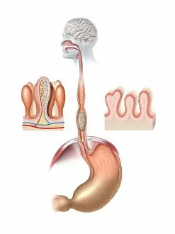
"Exploring the Intricate Network: Unveiling the Beauty of Capillaries" Delving into the depths of the human digestive system, capillaries emerge as delicate masterpieces


All Professionally Made to Order for Quick Shipping
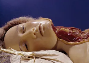
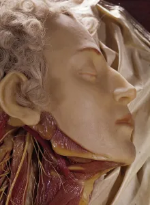
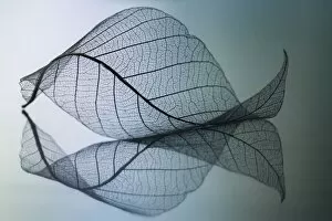
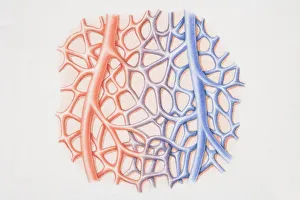
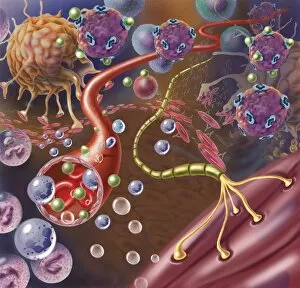
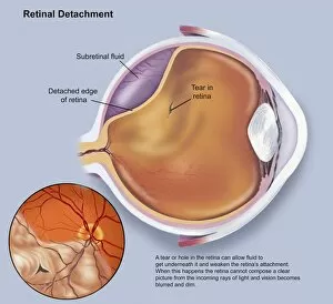
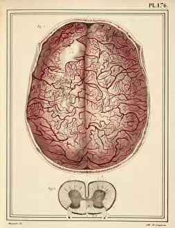
"Exploring the Intricate Network: Unveiling the Beauty of Capillaries" Delving into the depths of the human digestive system, capillaries emerge as delicate masterpieces. Like intricate artwork, they intricately weave through organs and tissues, ensuring vital nutrients reach every corner. In a mesmerizing display under scanning electron microscopy (SEM), kidney glomeruli reveal their captivating beauty. These tiny structures showcase an exquisite network that play a crucial role in filtering waste products from our blood. Moving on to another SEM marvel, we discover the thyroid gland's capillaries. Their intricate patterns resemble abstract art, highlighting their importance in regulating hormone production and metabolism. The kidney takes center stage once again as SEM captures its blood vessels with astonishing detail. The complexity and interconnectivity of these vessels are truly awe-inspiring, showcasing nature's remarkable design. Shifting our focus to the retina, SEM unveils a breathtaking view of its blood vessels. With their branching patterns resembling tree roots reaching for light, these capillaries nourish this essential part of our visual system. A false-color SEM image reveals a pericyte capillary on muscle—a true testament to both strength and fragility within our bodies. This microscopic wonder showcases how even at such small scales, life-sustaining processes occur seamlessly. Returning to kidney glomeruli—this time captured through light micrography—we witness their stunning architecture up close. These images remind us that beauty can be found even in places we least expect it. Tr@nsparences bring forth yet another dimension to understanding capillaries' significance—an artistic representation that merges science with creativity. Through this medium, we gain new perspectives on these vital conduits within us. Finally, a wax model depicting a man whose throat has been cut serves as a haunting reminder of how fragile our existence is without functioning capillaries. It underscores their indispensable role in sustaining life.