Mounted Print : Muscle fibres, SEM


![]()
Mounted Prints from Science Photo Library
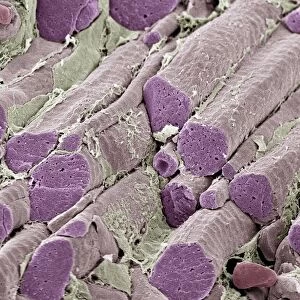
Muscle fibres, SEM
Skeletal muscle fibres. Coloured scanning electron micrograph (SEM) of a freeze-fractured bundle of skeletal (or striated) muscle fibres. The fracture planes across the fibres, and the removal of the sheath of connective tissue, has revealed the banding pattern that arises from the regular arrangement of the microscopic fibres (myofibrils) found in a muscle fibre. Skeletal muscle is under voluntary control. Magnification unknown
Science Photo Library features Science and Medical images including photos and illustrations
Media ID 6420424
© STEVE GSCHMEISSNER/SCIENCE PHOTO LIBRARY
Fibre Fibres Freeze Fracture Freeze Fractured Histological Histology Muscles Skeletal Striated Tissue Section Sectioned
14"x12" Mount with 12"x10" Print
Discover the intricacy of the human body with our Media Storehouse Mounted Photos featuring this captivating Scanning Electron Micrograph (SEM) of Skeletal Muscle Fibres by Science Photo Library. Witness the vibrant colours and intricate details of muscle fibres in this freeze-fractured bundle, revealing the complex structures that make up our skeletal system. Ideal for educational and scientific displays, these high-quality mounted photos bring the wonders of biology into your home or workplace.
Printed on 12"x10" paper and suitable for use in a 14"x12" frame (frame not included). Prints are mounted with card both front and back. Featuring a custom cut aperture to match chosen image. Professional 234gsm Fujifilm Crystal Archive DP II paper.
Photo prints supplied in custom cut card mount ready for framing
Estimated Image Size (if not cropped) is 27.7cm x 25.4cm (10.9" x 10")
Estimated Product Size is 35.6cm x 30.5cm (14" x 12")
These are individually made so all sizes are approximate
Artwork printed orientated as per the preview above, with landscape (horizontal) orientation to match the source image.
EDITORS COMMENTS
This print showcases the intricate beauty of skeletal muscle fibres, as captured through a scanning electron microscope (SEM). The coloured SEM image reveals a freeze-fractured bundle of these fibres, providing a unique glimpse into their inner structure. The removal of the connective tissue sheath and the fracture planes across the fibres have unveiled an awe-inspiring banding pattern. This pattern arises from the regular arrangement of microscopic myofibrils within each muscle fibre. These myofibrils play a crucial role in muscle contraction and are responsible for generating force and movement. Skeletal muscles, such as those depicted in this print, are under voluntary control. They enable us to perform various physical activities like walking, running, or lifting objects. Their health and proper functioning are essential for our overall well-being. This extraordinary image not only highlights the complexity and organization present within our bodies but also serves as a reminder of how remarkable human anatomy truly is. It offers viewers an opportunity to appreciate both the aesthetic appeal and scientific significance behind skeletal muscle fibres. Displayed by Science Photo Library, this print represents just one example of their vast collection that explores biology at its finest – showcasing nature's wonders on both macroscopic and microscopic scales.
MADE IN THE UK
Safe Shipping with 30 Day Money Back Guarantee
FREE PERSONALISATION*
We are proud to offer a range of customisation features including Personalised Captions, Color Filters and Picture Zoom Tools
SECURE PAYMENTS
We happily accept a wide range of payment options so you can pay for the things you need in the way that is most convenient for you
* Options may vary by product and licensing agreement. Zoomed Pictures can be adjusted in the Basket.