Mounted Print > Arts > Minimalist artwork > Monochrome artwork > Fine art
Mounted Print : Broken ankle, X-ray C017 / 7185


![]()
Mounted Prints from Science Photo Library
Broken ankle, X-ray C017 / 7185
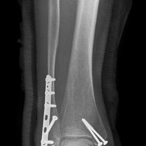
Broken ankle. X-ray of the lower leg of an 85 year old female with fractures at the ends of the tibia (shin bone, right) and fibula (calf bone, left). The fracture to the fibula is a spiral fracture, which is caused by a twisting force
Science Photo Library features Science and Medical images including photos and illustrations
Media ID 9342149
© SCIENCE PHOTO LIBRARY
Ankle Break Broken Diagnosis Diagnostic Eighties Elderly Fibula Fracture Fractured Fragments Injury Lower Leg Orthopaedics Orthopedics Patient Radiography Tibia X Ray Machine Xray Abnormal Injure Unhealthy
10"x8" Mount with 8"x6" Print
Explore the Media Storehouse range of Mounted Photos and bring the intricacies of medical science into your space. This captivating X-ray image, SCIENCE PHOTO LIBRARY's C017 / 7185, showcases the complex fractures at the ends of a senior woman's tibia and fibula in an 85-year-old female's broken ankle. Engage your audience with this thought-provoking visual representation of human anatomy and the healing process.
Printed on 8"x6" paper and suitable for use in a 10"x8" frame (frame not included). Prints are mounted with card both front and back. Featuring a custom cut aperture to match chosen image. Professional 234gsm Fujifilm Crystal Archive DP II paper.
Photo prints supplied in custom cut card mount ready for framing
Estimated Image Size (if not cropped) is 15.2cm x 18.6cm (6" x 7.3")
Estimated Product Size is 20.3cm x 25.4cm (8" x 10")
These are individually made so all sizes are approximate
Artwork printed orientated as per the preview above, with portrait (vertical) orientation to match the source image.
FEATURES IN THESE COLLECTIONS
> Arts
> Minimalist artwork
> Monochrome artwork
> Fine art
> Arts
> Minimalist artwork
> Monochrome artwork
> Monochrome paintings
EDITORS COMMENTS
This print from Science Photo Library showcases the X-ray of a broken ankle, belonging to an 85-year-old female patient. The image reveals fractures at both ends of her tibia (shin bone) and fibula (calf bone), with the latter displaying a distinctive spiral fracture resulting from a twisting force. The elderly woman's injury highlights the vulnerability that comes with age, as bones become more fragile and susceptible to damage. This diagnostic X-ray provides valuable insight into the severity of her condition, aiding medical professionals in formulating an appropriate treatment plan. The monochrome composition adds a sense of gravity to this visual representation of trauma within the human body. The fragmented bones are clearly visible, emphasizing the extent of damage caused by this unfortunate incident. Orthopaedics specialists may find particular interest in studying this image due to its relevance in diagnosing similar cases involving lower leg injuries among older adults. It serves as a reminder that even seemingly routine movements can result in severe consequences for individuals whose bones have weakened over time. Overall, this powerful photograph not only captures a moment frozen in time but also sheds light on the importance of proper care and attention towards our aging population's musculoskeletal health.
MADE IN THE UK
Safe Shipping with 30 Day Money Back Guarantee
FREE PERSONALISATION*
We are proud to offer a range of customisation features including Personalised Captions, Color Filters and Picture Zoom Tools
FREE COLORIZATION SERVICE
You can choose advanced AI Colorization for this picture at no extra charge!
SECURE PAYMENTS
We happily accept a wide range of payment options so you can pay for the things you need in the way that is most convenient for you
* Options may vary by product and licensing agreement. Zoomed Pictures can be adjusted in the Basket.