Mounted Print > Animals > Fishes > G > Grouper
Mounted Print : Diagram of lower leg illustrating muscle groups, nerves and veins


![]()
Mounted Prints from Fine Art Storehouse
Diagram of lower leg illustrating muscle groups, nerves and veins
Unleash your creativity and transform your space into a visual masterpiece!
Dorling Kindersley
Media ID 13560907
© This content is subject to copyright
Achilles Anatomy Ankle Artery Biology Biomedical Illustration Calf Diagram Foot Health Healthcare And Medicine Human Artery Human Body Human Joint Human Leg Human Muscle Lateral Muscle Flexor Digitorum Longus Human Body Part
10"x8" Mount with 8"x6" Print
Discover the intricacies of the lower leg with our exquisite Diagram of the Lower Leg from Dorling Kindersley's Fine Art Storehouse. This stunningly detailed mounted print showcases the complex network of muscles, nerves, and veins that make up this essential part of the human anatomy. Ideal for medical professionals, students, or anyone with a fascination for the human body, this beautifully presented piece adds a touch of sophistication and knowledge to any room. Bring the wonders of anatomy into your home or office with the Media Storehouse range of educational and fine art prints.
Printed on 8"x6" paper and suitable for use in a 10"x8" frame (frame not included). Prints are mounted with card both front and back. Featuring a custom cut aperture to match chosen image. Professional 234gsm Fujifilm Crystal Archive DP II paper.
Photo prints supplied in custom cut card mount ready for framing
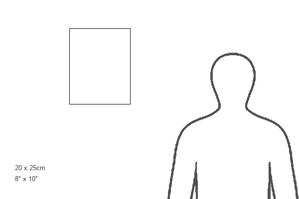
Estimated Image Size (if not cropped) is 13.5cm x 20.3cm (5.3" x 8")
Estimated Product Size is 20.3cm x 25.4cm (8" x 10")
These are individually made so all sizes are approximate
Artwork printed orientated as per the preview above, with portrait (vertical) orientation to match the source image.
FEATURES IN THESE COLLECTIONS
> Fine Art Storehouse
> Photo Libraries
> Dorling Kindersley Prints
> Animals
> Fishes
> G
> Grouper
EDITORS COMMENTS
This print by Dorling Kindersley showcases a detailed diagram of the lower leg, providing an insightful glimpse into the intricate musculoskeletal system. The image beautifully illustrates various muscle groups, nerves, and veins that play a vital role in our everyday movements. From the Achilles tendon to the flexor digitorum longus and hallucis longus muscles, every anatomical feature is meticulously depicted with precision and clarity. The crural intermuscular septum divides different compartments of the leg while highlighting their distinct functions. The vertical composition of this biomedical illustration allows for easy comprehension as it guides us through each element step by step. Its vibrant colors enhance visual appeal without compromising scientific accuracy. With no people present in the frame, this artwork solely focuses on unraveling the complexity of human anatomy. It serves as an invaluable resource for healthcare professionals, students studying biology or medicine, or anyone intrigued by understanding their own body better. Dorling Kindersley's expertise shines through in this exceptional piece that seamlessly combines artistry with educational value. This print is not only aesthetically pleasing but also serves as a testament to how art can contribute to our knowledge and appreciation of the human body's remarkable intricacies.
MADE IN THE UK
Safe Shipping with 30 Day Money Back Guarantee
FREE PERSONALISATION*
We are proud to offer a range of customisation features including Personalised Captions, Color Filters and Picture Zoom Tools
SECURE PAYMENTS
We happily accept a wide range of payment options so you can pay for the things you need in the way that is most convenient for you
* Options may vary by product and licensing agreement. Zoomed Pictures can be adjusted in the Basket.