
Human Joint Collection
"Exploring the Intricacies of Human Joints: From Shoulders to Knees and Everything In Between" In this captivating journey through the human body


All Professionally Made to Order for Quick Shipping
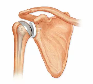
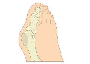
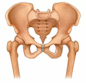
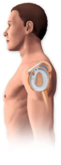
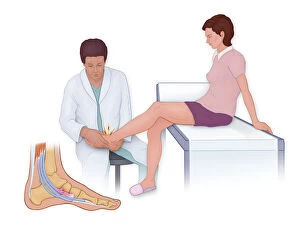
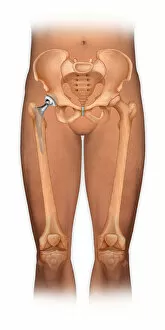
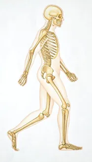
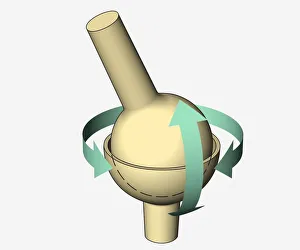
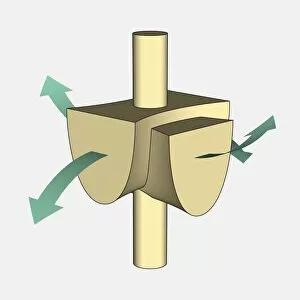
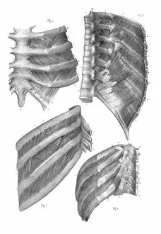
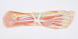
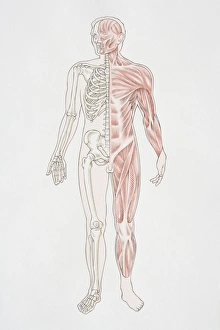
"Exploring the Intricacies of Human Joints: From Shoulders to Knees and Everything In Between" In this captivating journey through the human body, we delve into the fascinating world of joints. Starting with an anterior view of a total shoulder joint repair, we witness the intricate process of restoring mobility and function. Amidst the enchanting scene of a teen girl surrounded by vinyl records and a record player, we are reminded that our joints enable us to embrace life's melodies while sitting comfortably on the floor. The hand, an incredible marvel in itself, showcases its dexterity and power. Its delicate yet strong structure allows us to create art, express love, and accomplish remarkable feats. Moving further up towards our most enigmatic feature - the human skull. Within its complex framework lies protection for our precious brain as well as various joints that allow for movement and expression. Transitioning down to one of our weight-bearing wonders - the knee joint. An engraving from 1866 reveals its intricate anatomy while highlighting how it has evolved over time. As we explore deeper into anatomical engravings showcasing both male and female bodies, we encounter another masterpiece –the endocrine system. This vital network regulates countless bodily functions including those related to joint health. Returning back down towards our pelvises with hip bones intact, we observe their normal anterior view – a testament to stability and grace in motion. However, not all journeys are without obstacles; arthritis takes center stage as femoral heads display osteophytes in an anterior view affected by this condition. Yet even amidst adversity, these resilient structures continue supporting us every step of the way. Finally, witnessing surgical intervention using a bovie tool reminds us that medical advancements can restore damaged joints like displaced patellar knees – offering hope for improved quality of life. From shoulder repairs to artistic endeavors encompassed within vinyl records or engraved illustrations dating back centuries ago – each caption unveils the intricate beauty and functionality of human joints.