Greetings Card > Science > SEM
Greetings Card : Stomach wall, SEM


![]()
Cards from Science Photo Library
Stomach wall, SEM
Stomach wall. Coloured scanning electron micrograph (SEM) of a section through a freeze- fractured fold in the stomach wall. The stomach is lined by the mucosa (purple), whose glands secrete gastric juices into the stomach lumen (cavity, top). At the base of the mucosa is the muscularis mucosae (light brown), a thin layer of smooth muscle which provides local movements and folding of the mucosa. The submucosa (beige) lies beneath the muscularis mucosae. It is a layer of loose connective tissue that contains blood vessels and nerves (not seen). Beneath the submucosa is the muscularis (dark red), a layer of smooth muscle. Magnification: x50 at 6x7cm size
Science Photo Library features Science and Medical images including photos and illustrations
Media ID 6450107
© STEVE GSCHMEISSNER/SCIENCE PHOTO LIBRARY
Alimentary Canal Connective Digestion Digestive System Fold Folded Folding Fracture Freeze Fractured Gastric Histology Layers Magnified Image Microscopic Photos Mucosa Mucosal Muscular Smooth Stomach Sub Mucosa Subjects Tissue Tract Wall Section Sectioned
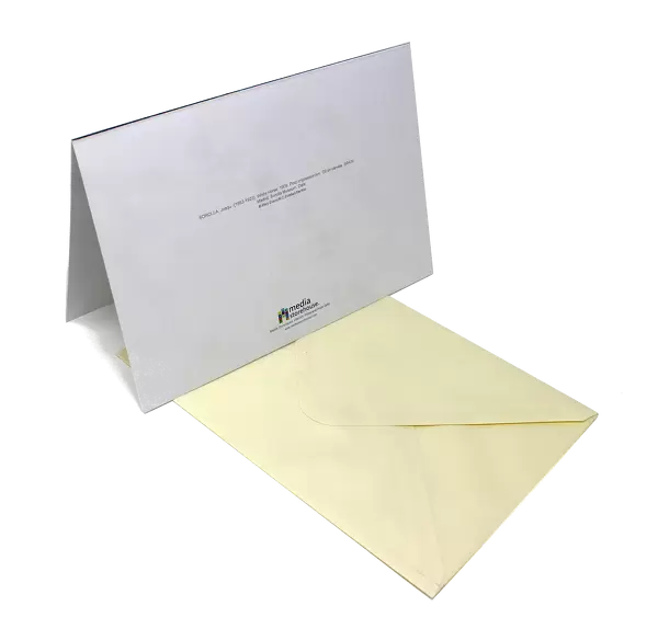
Greetings Card Large (A4)
Discover the wonders of the human body with our selection of unique Science-themed greeting cards from Media Storehouse. This card features a captivating coloured Scanning Electron Micrograph (SEM) image of a section through a freeze-fractured fold in the stomach wall, titled "Stomach wall, SEM" by Science Photo Library. Impress your loved ones with this stunning representation of the complex structures that make up our bodies. Perfect for birthdays, anniversaries, or just to brighten someone's day, these cards are a thoughtful and scientifically-inspired way to show you care.
Create your own large greetings card. Size when folded is A4 (21x30cm or 8.3x11.7 inches)
Greetings Cards suitable for Birthdays, Weddings, Anniversaries, Graduations, Thank You and much more
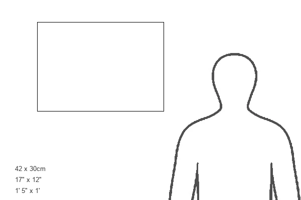
Estimated Image Size (if not cropped) is 21cm x 29.7cm (8.3" x 11.7")
Estimated Product Size is 42cm x 29.7cm (16.5" x 11.7")
These are individually made so all sizes are approximate
Artwork printed orientated as per the preview above, with portrait (vertical) orientation to match the source image.
EDITORS COMMENTS
This print showcases the intricate beauty of the stomach wall, as seen through a scanning electron microscope (SEM). The image reveals a freeze-fractured fold in the stomach wall, providing us with an up-close and detailed view of its anatomy. The mucosa, depicted in vibrant purple hues, lines the inner surface of the stomach. This layer contains glands that secrete gastric juices into the cavity of the stomach. Just beneath it lies the muscularis mucosae, represented by light brown tones. This thin layer of smooth muscle facilitates local movements and folding of the mucosa. In this magnified image, we can also observe the submucosa in beige shades. Composed of loose connective tissue, it houses blood vessels and nerves that support proper functioning but are not visible here. Further down is the muscularis itself, portrayed in dark red colors. This robust layer consists of smooth muscle responsible for propelling food along during digestion. Overall, this SEM photograph offers a glimpse into a healthy and normal sectioned portion of our digestive system's alimentary canal—the stomach. Its vivid colors and intricate layers provide valuable insights into how our bodies function on a microscopic level while highlighting both its complexity and elegance.
MADE IN THE UK
Safe Shipping with 30 Day Money Back Guarantee
FREE PERSONALISATION*
We are proud to offer a range of customisation features including Personalised Captions, Color Filters and Picture Zoom Tools
SECURE PAYMENTS
We happily accept a wide range of payment options so you can pay for the things you need in the way that is most convenient for you
* Options may vary by product and licensing agreement. Zoomed Pictures can be adjusted in the Basket.