Glass Frame > Popular Themes > Human Body
Glass Frame : Lung alveoli


![]()
Mounted Prints from Science Photo Library
Lung alveoli
Lung alveoli. Light micrograph of a section through normal human lung tissue showing alveoli. These are tiny air sacs where most of the diffusion of gases to and from the blood occurs. They have thin walls (red) lined with capillaries (not seen). Inhaled oxygen diffuses into the blood in these capillaries, and carbon dioxide is released and exhaled. The oxygenated blood travels to the heart where it is pumped around the body, releasing its oxygen and picking up carbon dioxide before returning to the lungs. Magnification unknown
Science Photo Library features Science and Medical images including photos and illustrations
Media ID 6450093
© CNRI/SCIENCE PHOTO LIBRARY
Alveoli Alveolus Breathing Histological Histology Lung Lungs Microscope Net Work Respiration Respiratory System Slide Light Micrograph Section Sectioned
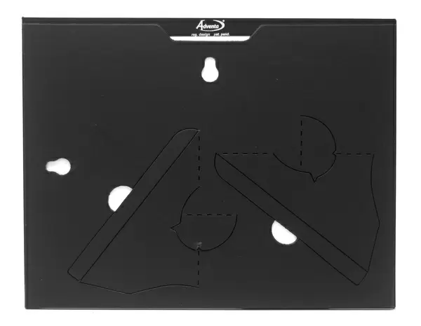
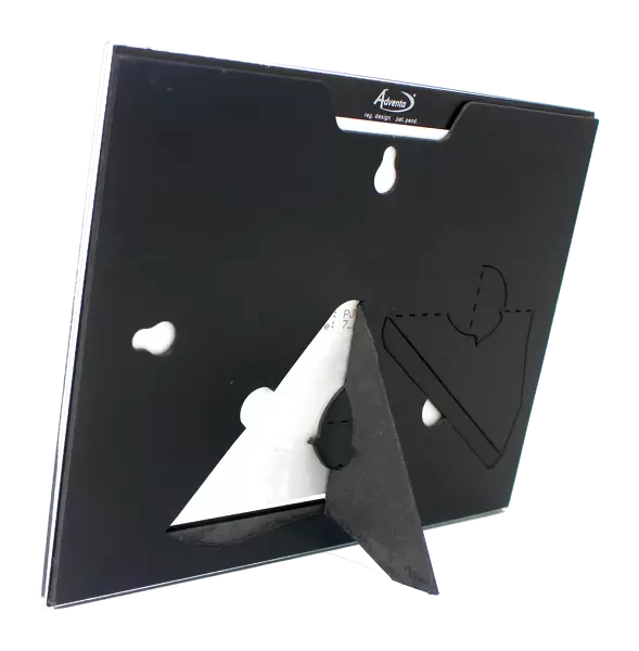
7"x5" Glass Mount
Wall mounted or free-standing, these black edged glass frames feature a smooth chamfered edge and a stylish black border (on back face of the glass). Manufactured from 4mm thick glass, Glass Mounts are a durable, professional way of displaying and protecting your prints. Your 7x5 print is slotted into the back of the frame so can easily be changed if needed.
Tempered Glass Mounts are ideal for wall display, plus the smaller sizes can also be used free-standing via an integral stand
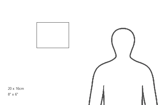
Estimated Image Size (if not cropped) is 17.7cm x 12.7cm (7" x 5")
Estimated Product Size is 20.3cm x 16.2cm (8" x 6.4")
These are individually made so all sizes are approximate
Artwork printed orientated as per the preview above, with landscape (horizontal) orientation to match the source image.
EDITORS COMMENTS
This print from Science Photo Library showcases the intricate beauty of lung alveoli, the tiny air sacs within our lungs. The image, captured using a light microscope, provides a glimpse into the remarkable network that enables efficient gas exchange in our respiratory system. The vibrant red walls of these alveoli are incredibly thin and delicately lined with capillaries, although not visible in this particular micrograph. It is within these microscopic structures that the crucial diffusion of gases takes place – oxygen entering the bloodstream while carbon dioxide is released and exhaled. As we breathe in, oxygen molecules effortlessly diffuse through these thin walls into the surrounding capillaries. This freshly oxygenated blood then embarks on an incredible journey to our heart where it will be pumped throughout our body. Along its path, it releases its life-sustaining cargo of oxygen and picks up carbon dioxide waste before returning to the lungs for another round. The magnification used to capture this mesmerizing image remains unknown; however, what is evident is how intricately designed and interconnected our bodies truly are. This histological masterpiece serves as a reminder of just how vital each breath we take truly is for sustaining life itself.
MADE IN THE UK
Safe Shipping with 30 Day Money Back Guarantee
FREE PERSONALISATION*
We are proud to offer a range of customisation features including Personalised Captions, Color Filters and Picture Zoom Tools
SECURE PAYMENTS
We happily accept a wide range of payment options so you can pay for the things you need in the way that is most convenient for you
* Options may vary by product and licensing agreement. Zoomed Pictures can be adjusted in the Basket.