
Respiratory System Collection
The respiratory system is a complex network of organs and tissues that work together to ensure our bodies receive the oxygen they need


All Professionally Made to Order for Quick Shipping
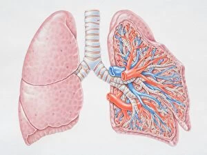
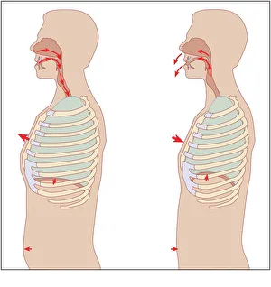
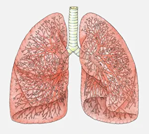
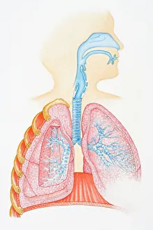
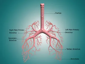
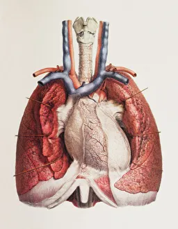
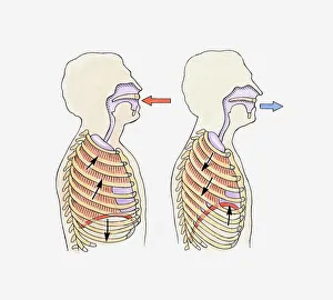
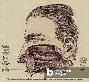
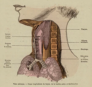
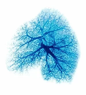
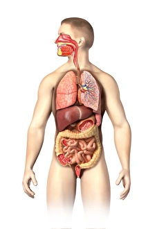
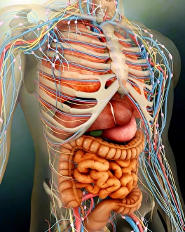
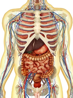
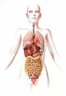
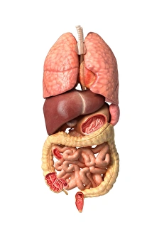
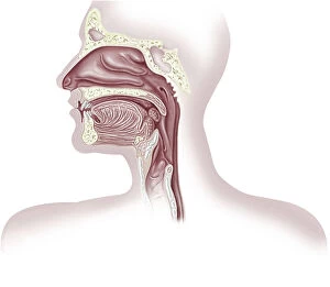
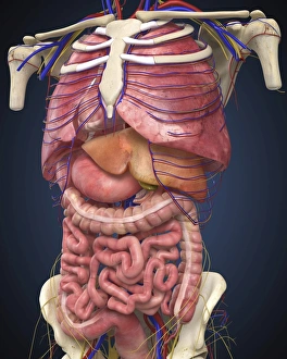
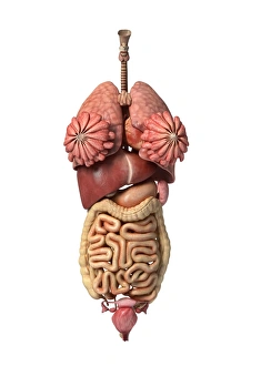
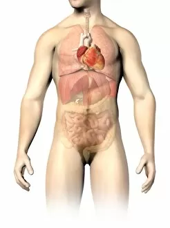
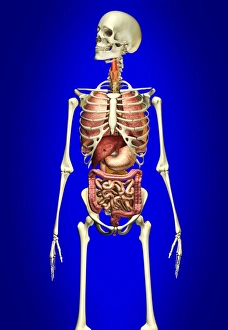
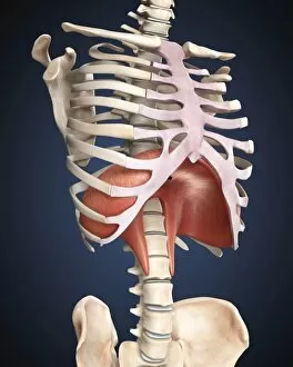
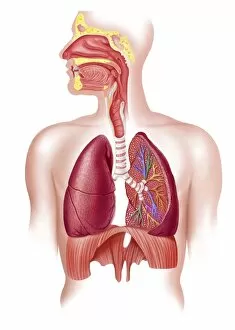
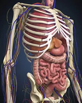
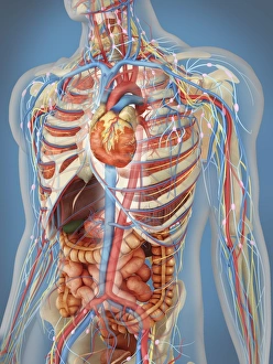
The respiratory system is a complex network of organs and tissues that work together to ensure our bodies receive the oxygen they need. From the mechanics of respiration to the intricate anatomy, this caption will take you on a journey through this vital system. Starting with a diagram, we can see how each component fits into place. The oral and nasal cavities act as entry points for air, leading to the larynx, trachea, bronchus, and finally reaching the lungs. This illustration showcases just how interconnected these structures are. Moving closer into the lungs, we can observe their incredible blood supply in another diagram. It's fascinating to see how every breath we take allows oxygen to be transported throughout our bodies via this intricate network. In times of medical intervention or emergency situations, an oxygen mask becomes essential. This simple device helps deliver concentrated oxygen directly into our respiratory system when needed most. Zooming in further reveals the detailed anatomy of bronchi and bronchial tubes. These branching passageways play a crucial role in ensuring air reaches all parts of our lungs efficiently. The heart and lungs have an inseparable relationship as they work harmoniously together to keep us alive. Their close proximity is evident in various illustrations showcasing their connection. A front view diagram gives us an inside look at the structure of our lungs themselves – two remarkable organs responsible for gas exchange within tiny sacs called alveoli. Examining even smaller details under scanning electron microscopy (SEM), we discover the delicate lining of our trachea – yet another testament to nature's intricacy. Understanding how breathing occurs involves observing movements such as diaphragm contraction during inhalation and relaxation during exhalation. An illustration captures these actions perfectly. To assess lung function accurately or diagnose potential issues, lung function tests become invaluable tools for healthcare professionals seeking comprehensive insights into respiratory health. An X-ray image offers a glimpse beneath our skin's surface – revealing any abnormalities or conditions that may be affecting our lungs.