Glass Frame : Chloroplast in cell of pea plant


![]()
Mounted Prints from Science Photo Library
Chloroplast in cell of pea plant
Coloured transmisson electron micrograph of a chloroplast (green) sitting in the cytoplasm of a pea plant Pisum sativum. The chloroplast is the site of photosynthesis where carbohydrates are obtained from carbon dioxide using the energy from sunlight. The chloroplast is bound by a double membrane. Internally, it consists of stacks of flattened membranes called grana (threadlike) suspended in a matrix of hydrophilic proteins. The grana contain the chlorophyll pigments and are the sites of light reactions during photosynthesis. The orange at left is part of a nucleus and the orange strip at right is the cell wall. Magnification: x1680 at 6x4.5cm size
Science Photo Library features Science and Medical images including photos and illustrations
Media ID 6293691
© DR JEREMY BURGESS/SCIENCE PHOTO LIBRARY
Botanical Science Chloroplast Chloroplasts Electron Micrograph Grana Photosynthesis Pisum Sativum Plant Structure Plastid Plastids Starch Transmission
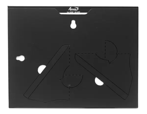
7"x5" Glass Mount
Wall mounted or free-standing, these black edged glass frames feature a smooth chamfered edge and a stylish black border (on back face of the glass). Manufactured from 4mm thick glass, Glass Mounts are a durable, professional way of displaying and protecting your prints. Your 7x5 print is slotted into the back of the frame so can easily be changed if needed.
Tempered Glass Mounts are ideal for wall display, plus the smaller sizes can also be used free-standing via an integral stand
Estimated Image Size (if not cropped) is 12.7cm x 17.7cm (5" x 7")
Estimated Product Size is 16.2cm x 20.3cm (6.4" x 8")
These are individually made so all sizes are approximate
Artwork printed orientated as per the preview above, with portrait (vertical) orientation to match the source image.
EDITORS COMMENTS
This print showcases the intricate beauty of a chloroplast within a cell of a vibrant pea plant. In this coloured transmission electron micrograph, the chloroplast is depicted as a striking shade of green, contrasting against the surrounding cytoplasm. Serving as the epicenter of photosynthesis, this vital organelle converts carbon dioxide into carbohydrates using sunlight as its energy source. Enclosed by a double membrane, the chloroplast reveals its internal structure through stacks of flattened membranes known as grana. These threadlike structures are suspended in a matrix composed of hydrophilic proteins. It is within these grana that chlorophyll pigments reside and where light reactions occur during photosynthesis. The orange hue on the left side represents part of a nucleus while an orange strip on the right signifies the presence of the cell wall. With an impressive magnification level at 1680x and measuring 6x4.5cm in size, this image provides an extraordinary glimpse into botanical science and plant structure. Through this visually stunning representation captured by Science Photo Library, we gain insight into the complex world within plants - specifically highlighting how chloroplasts play an essential role in sustaining life through their remarkable ability to harness sunlight's power for carbohydrate synthesis.
MADE IN THE UK
Safe Shipping with 30 Day Money Back Guarantee
FREE PERSONALISATION*
We are proud to offer a range of customisation features including Personalised Captions, Color Filters and Picture Zoom Tools
SECURE PAYMENTS
We happily accept a wide range of payment options so you can pay for the things you need in the way that is most convenient for you
* Options may vary by product and licensing agreement. Zoomed Pictures can be adjusted in the Basket.