Fraud Blocker Skip to main content

Scanning Electron Microscope Collection (page 8)

"Unveiling the Hidden World

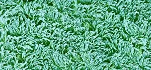
Background imageScanning Electron Microscope Collection: Pipistrelle bat ear, SEM

Pipistrelle bat ear, SEM
Pipistrelle bat ear. Coloured scanning electron micrograph (SEM) of the ear of a pipistrelle bat (Pipistrellus pipistrellus)

Background imageScanning Electron Microscope Collection: Turtle ants head, SEM

Turtle ants head, SEM
Turtle ants head. Coloured scanning electron micrograph (SEM) of the head of a soldier turtle ant (Cephalotes sp.) from the Amazonian rainforest. One of its compound eyes (green) is at left

Background imageScanning Electron Microscope Collection: Female mosquito head, SEM

Female mosquito head, SEM
Female mosquito head. Coloured scanning electron micrograph (SEM) of the head of a female mosquito (family Culicidae). The mosquitos head is dominated by its large compound eyes (pink)

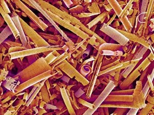
Background imageScanning Electron Microscope Collection: Metal shavings

Metal shavings. Coloured scanning electron micrograph (SEM) of metal shavings taken from a key cutter s

Background imageScanning Electron Microscope Collection: Komodo dragon tooth, SEM

Komodo dragon tooth, SEM
Komodo dragon tooth, coloured scanning electron micrograph (SEM). This curved serrated tooth is designed to slice, cut and tear meat, but not to chew it

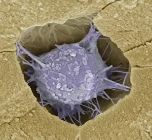
Background imageScanning Electron Microscope Collection: Osteoblast bone cell, SEM

Osteoblast bone cell, SEM
Osteoblast bone cell. Coloured scanning electron micrograph (SEM) of an osteoblast (centre) in bone tissue. Osteoblasts are primarily found in regions of new bone growth

Background imageScanning Electron Microscope Collection: Ant, SEM

Ant, SEM
Ant (family Formicidae), coloured scanning electron micrograph (SEM). One of its compound eyes (black) is seen, as well as two long antennae that are mounted on the head between the eyes

Background imageScanning Electron Microscope Collection: Mitochondria, SEM

Mitochondria, SEM
Mitochondria. Coloured scanning electron micrograph (SEM) of mitochondria (red) in a kidney cell. Mitochondria are a type of organelle found in the cytoplasm of eukaryotic cells

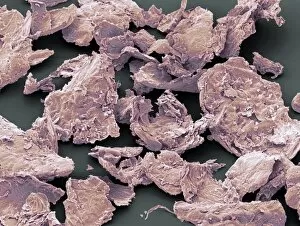
Background imageScanning Electron Microscope Collection: Flakes of dead skin, SEM

Flakes of dead skin, SEM
Flakes of dead skin. Coloured scanning electron micrograph (SEM) of dead skin from the scalp (dandruff), a condition where there is an excessive shedding of dead skin cells

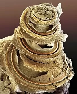
Background imageScanning Electron Microscope Collection: Organ of Corti, inner ear, SEM

Organ of Corti, inner ear, SEM
Organ of Corti. Coloured scanning electron micrograph (SEM) of the organ of Corti from the cochlea of the inner ear. The bony capsule that encloses the organ has been removed to reveal the organs

Background imageScanning Electron Microscope Collection: Chickweed pollen grain, SEM

Chickweed pollen grain, SEM
Chickweed pollen grain. Coloured scanning electron micrograph (SEM) of a pollen grain (green) from a chickweed flower (Stellaria media)

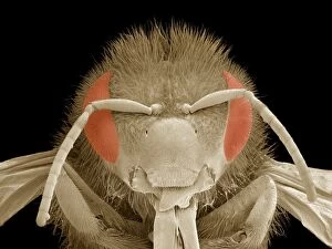
Background imageScanning Electron Microscope Collection: Honey bee head, SEM

Honey bee head, SEM
Honey bee head. Coloured scanning electron micrograph (SEM) of the head of a honey bee (Apis sp.). The compound eyes (red) are either side of the antennae

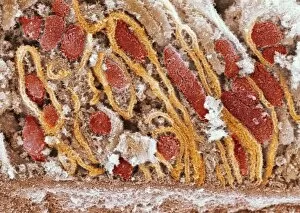
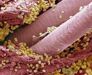
Background imageScanning Electron Microscope Collection: Lymphocytes in hair follicle, SEM

Lymphocytes in hair follicle, SEM
Lymphocytes in hair follicle, coloured scanning electron micrograph (SEM). Large numbers of lymphocytes (yellow) are seen in inflamed and necrotic tissue at the base of hair shafts (red)

Background imageScanning Electron Microscope Collection: Bacillus subtilis bacteria, SEM

Bacillus subtilis bacteria, SEM
Bacillus subtilis. Coloured scanning electron micrograph (SEM) of Bacillus subtilis bacteria. B. subtilis, or hay bacillus, is an aerobic, gram-positive bacillus(rod-shaped bacterium)

Background imageScanning Electron Microscope Collection: Lactobacillus bacteria, SEM

Lactobacillus bacteria, SEM
Lactobacillus sp. bacteria, coloured scanning electron micrograph (SEM). Lactobacillus bacteria are probiotic bacteria, which are useful for human and animal health as they produce bacteriocins

Background imageScanning Electron Microscope Collection: Starch grains, SEM

Starch grains, SEM
Starch grains. Coloured scanning electron micrograph (SEM) of starch grains (orange) in a black-eyed pea (Vigna sp.). Starch is synthesised from sucrose

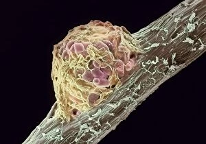
Background imageScanning Electron Microscope Collection: Root nodule

Root nodule. Coloured scanning electron micrograph (SEM) of a root nodule on a pea plant (Pisum sativum) caused by the nitrogen-fixing soil bacteria Rhizobium leguminosarum

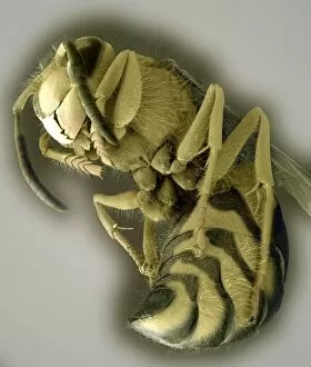
Background imageScanning Electron Microscope Collection: Common wasp, SEM

Common wasp, SEM
Common wasp (Vespula vulgaris), coloured scanning electron micrograph (SEM). These wasps are social insects. They live in underground nests of up to around 10, 000 workers

Background imageScanning Electron Microscope Collection: Bamboo shoot weevil, SEM

Bamboo shoot weevil, SEM
Bamboo shoot weevil (Otidognathus davidis), coloured scanning electron micrograph (SEM). This insect is a pest of bamboo shoots, and is widespread in the Far East

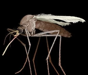
Background imageScanning Electron Microscope Collection: Scanning Electron Micrograph (SEM): Mosquito, Magnification x 35 (A4 size: 29. 7 cm width)

Scanning Electron Micrograph (SEM): Mosquito, Magnification x 35 (A4 size: 29. 7 cm width)
LRDS-84 Mosquito Scanning Electron Micrograph (SEM) Culex pipiens Magnification x 35 (A4 size: 29.7 cm width) Coloured by hand to enhance natural features

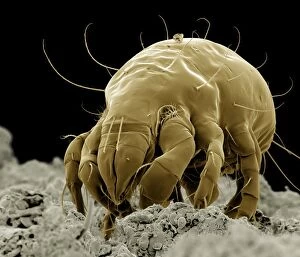
Background imageScanning Electron Microscope Collection: Scanning Electron Micrograph (SEM): Flour Mite, Magnification x 500 (A4 size: 29. 7 cm width)

Scanning Electron Micrograph (SEM): Flour Mite, Magnification x 500 (A4 size: 29. 7 cm width)
LRDS-58 Flour Mite Scanning Electron Micrograph (SEM) Acarus siro Magnification x 500 (A4 size: 29.7 cm width) Coloured by hand to enhance natural features

Background imageScanning Electron Microscope Collection: Scanning Electron Micrograph (SEM): Cat Flea, Magnification x250 (A4 size: 29. 7 cm width)

Scanning Electron Micrograph (SEM): Cat Flea, Magnification x250 (A4 size: 29. 7 cm width)
LRDS-54 Cat Flea Scanning Electron Micrograph (SEM) Ctenocephalides felis Magnification x250 (A4 size: 29.7 cm width) Coloured by hand to enhance natural features

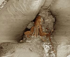
Background imageScanning Electron Microscope Collection: Scanning Electron Micrograph (SEM): Chigoe Flea, Magnification x 200 (A4 size: 29. 7 cm width)

Scanning Electron Micrograph (SEM): Chigoe Flea, Magnification x 200 (A4 size: 29. 7 cm width)
LRDS-137 Chigoe Flea Scanning Electron Micrograph (SEM) Tunga penetrans Magnification x 200 (A4 size: 29.7 cm width) Coloured by hand to enhance natural features

Background imageScanning Electron Microscope Collection: Scanning Electron Micrograph (SEM): Springtail, Order Collembola; Magnification x 150 (A4 size)

Scanning Electron Micrograph (SEM): Springtail, Order Collembola; Magnification x 150 (A4 size: 29. 7 cm width)
LRDS-122 Springtail, Order Collembola Scanning Electron Micrograph (SEM) Magnification x 150 (A4 size: 29.7 cm width) Coloured by hand to enhance natural features

Background imageScanning Electron Microscope Collection: Chrysanthemum, CT scan image

Chrysanthemum, CT scan image
CT Scan image of a Chrysanthemum

Background imageScanning Electron Microscope Collection: Syracosphaera anthos

Syracosphaera anthos
Coccosphere from the Western Mediterranean. False coloured to show the shell is formed of inner and outer layers of coccoliths with very different structure

Background imageScanning Electron Microscope Collection: Colorized image of HIV-infected H9 T-cell

Colorized image of HIV-infected H9 T-cell

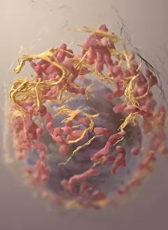
Background imageScanning Electron Microscope Collection: 3D structure of melanoma cell

3D structure of melanoma cell
3D structure of a melanoma cell derived by ion abrasion scanning electron microscopy

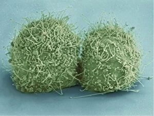
Background imageScanning Electron Microscope Collection: Scanning electron micrograph of just-divided HeLa cells

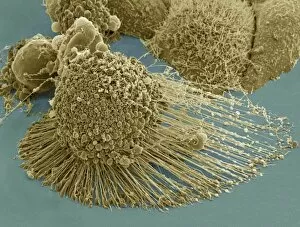
Scanning electron micrograph of just-divided HeLa cells. Zeiss Merlin HR-SEM

Background imageScanning Electron Microscope Collection: A colorized scanning electron micrograph of a white blood cell eating MRSA

A colorized scanning electron micrograph of a white blood cell eating MRSA
A colorized scanning electron micrograph of a white blood cell eating an antibiotic resistant strain of Staphylococcus aureus bacteria, commonly known as MRSA

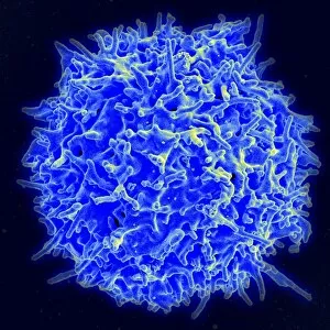
Background imageScanning Electron Microscope Collection: Scanning electron micrograph of a human T cell

Scanning electron micrograph of a human T cell
Scanning electron micrograph of a human T lymphocyte (also called a T cell) from the immune system of a healthy donor

Background imageScanning Electron Microscope Collection: Scanning electron micrograph of a human neutrophil ingesting MRSA

Scanning electron micrograph of a human neutrophil ingesting MRSA

Background imageScanning Electron Microscope Collection: Scanning electron micrograph of Staphylococcus and a dead human neutrophil

Scanning electron micrograph of Staphylococcus and a dead human neutrophil
Scanning electron micrograph of methicillin-resistant Staphylococcus aureus and a dead human neutrophil

Background imageScanning Electron Microscope Collection: Colorized scanning electron micrograph of a T lymphocyte

Colorized scanning electron micrograph of a T lymphocyte

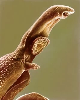
Background imageScanning Electron Microscope Collection: Scanning electron micrograph of a schistosome parasite

Scanning electron micrograph of a schistosome parasite, which enters the body through the skin of persons coming in contact with infested waters. The adult worm lives in the veins of its host

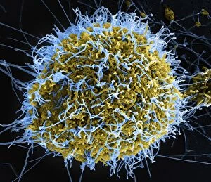
Background imageScanning Electron Microscope Collection: Colorized scanning electron micrograph of filamentous Ebola virus particles

Colorized scanning electron micrograph of filamentous Ebola virus particles (blue) budding from a chronically infected VERO E6 cell (yellow-green)

Background imageScanning Electron Microscope Collection: Scanning electron micrograph of an apoptotic HeLa cell

Scanning electron micrograph of an apoptotic HeLa cell. Zeiss Merlin HR-SEM

Background imageScanning Electron Microscope Collection: Schistosome fluke, SEM

Schistosome fluke, SEM
Schistosome fluke. Coloured scanning electron micrograph (SEM) of a schistosome (Schistosoma sp.) fluke worm, a cause of schistosomiasis in humans

Background imageScanning Electron Microscope Collection: Biology, Bacteria, Cocci seen through scanning electron microscope

Biology, Bacteria, Cocci seen through scanning electron microscope
Biology - Bacteria - Cocci seen through the scanning electron microscope

Background imageScanning Electron Microscope Collection: Blood clot, SEM C016 / 9751

Blood clot, SEM C016 / 9751
Blood clot, coloured scanning electron micrograph (SEM). Red blood cells (erythrocytes) are trapped within a fibrin protein mesh (beige)

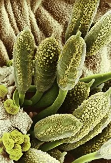
Background imageScanning Electron Microscope Collection: Carnation flower pistil, SEM C016 / 9762

Carnation flower pistil, SEM C016 / 9762
Carnation (Dianthus sp.) pistil, coloured scanning electron micrograph (SEM). Pistils hold a plants female reproductive structures

Background imageScanning Electron Microscope Collection: Blood clot, SEM C016 / 9746

Blood clot, SEM C016 / 9746
Blood clot, coloured scanning electron micrograph (SEM). Red blood cells (erythrocytes) are trapped within a fibrin protein mesh (beige)

Background imageScanning Electron Microscope Collection: Carnation flower pistil, SEM C016 / 9759

Carnation flower pistil, SEM C016 / 9759
Carnation (Dianthus sp.) pistil, coloured scanning electron micrograph (SEM). Pistils hold a plants female reproductive structures

Background imageScanning Electron Microscope Collection: Rust fungus on a rose leaf, SEM C017 / 7132

Rust fungus on a rose leaf, SEM C017 / 7132
Rust fungus on a rose leaf. Coloured scanning electron micrograph (SEM) of rust fungus (Phragmidium sp.) spores emerging from a rose (Rosa sp.) leaf (brown)

Background imageScanning Electron Microscope Collection: Blood clot, SEM C016 / 9749

Blood clot, SEM C016 / 9749
Blood clot, coloured scanning electron micrograph (SEM). Red blood cells (erythrocytes) are trapped within a fibrin protein mesh (beige)

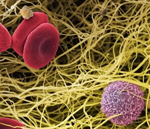
Background imageScanning Electron Microscope Collection: Blood clot, SEM C017 / 7141

Blood clot, SEM C017 / 7141
Blood clot, coloured scanning electron micrograph (SEM). Red blood cells (erythrocytes) and a white blood cell (purple) trapped within a fibrin protein mesh (cream)

Background imageScanning Electron Microscope Collection: Trachea lining, SEM C016 / 9756

Trachea lining, SEM C016 / 9756
Trachea lining. Coloured scanning electron micrograph (SEM) of the lining of the trachea (wind pipe). The trachea links the larynx to the lungs


All Professionally Made to Order for Quick Shipping

"Unveiling the Hidden World: Exploring with a Scanning Electron Microscope" The scanning electron microscope (SEM) is an incredible tool that allows us to delve into the microscopic realm, revealing intricate details and unlocking secrets of the unseen. With its high magnification capabilities, it offers a glimpse into fascinating aspects of life and nature. In one captivating image, we witness the beauty of gender determination as X and Y chromosomes are captured under the SEM's lens. The intricate patterns and structures within these chromosomes showcase the complexity of our genetic makeup. Moving on to another astonishing discovery, a Scanning Electron Micrograph presents us with an up-close view of a Praying Mantis at 30 times magnification. Every tiny detail becomes visible, from its delicate limbs to its mesmerizing eyes – reminding us of nature's remarkable diversity. Shifting focus to plant life, lavender pollen grains come alive in another SEM image. These minuscule particles take center stage as their unique shapes and textures become apparent under intense scrutiny. Zooming in even further reveals a Tardigrade or 'Water Bear' at an astounding 1250 times magnification. This resilient creature captures our imagination as we explore its alien-like features through this powerful imaging technique. Delving deeper into biological wonders, T lymphocytes battling cancer cells are showcased in yet another breathtaking SEM capture. This visual representation highlights the ongoing fight within our bodies against diseases like cancer – emphasizing both hope and resilience. Calcareous phytoplankton takes center stage next as we uncover their intricate forms through SEM imagery. These microscopic organisms play crucial roles in marine ecosystems while leaving us awe-inspired by their stunningly detailed structures. Transitioning to neural marvels, nerve cells reveal their intricacy under the watchful eye of an SEM. Their branching extensions create complex networks that enable communication throughout our bodies – showcasing nature's ingenuity at work. Venturing into insect realms once again, a fruit fly is immortalized under the SEM's lens.