Fraud Blocker Skip to main content

Scanning Electron Microscope Collection (page 4)

"Unveiling the Hidden World

Background imageScanning Electron Microscope Collection: Mouse malaria parasite, SEM

Mouse malaria parasite, SEM
Mouse malaria parasite. Coloured scanning electron micrograph (SEM) of a Plasmodium berghei protozoan (yellow) and red blood cells. P. berghei is the parasite that causes malaria in mice

Background imageScanning Electron Microscope Collection: Orchid petal, SEM

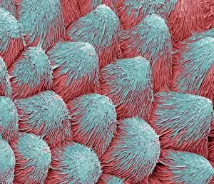
Orchid petal, SEM
Orchid (Phalaenopsis sp.) petal, coloured scanning electron micrograph (SEM). Magnification: x450 when printed at 10 centimetres wide

Background imageScanning Electron Microscope Collection: Stem cell, SEM

Stem cell, SEM
Stem cell, coloured scanning electron micrograph (SEM). Stem cells can differentiate into any other cell type. There are three main types of mammalian stem cell: embryonic stem cells

Background imageScanning Electron Microscope Collection: Stem cells, SEM

Stem cells, SEM
Stem cells, coloured scanning electron micrograph (SEM). Stem cells can differentiate into any other cell type. There are three main types of mammalian stem cell: embryonic stem cells

Background imageScanning Electron Microscope Collection: Mycobacterium chelonae bacteria, SEM

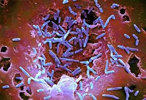
Mycobacterium chelonae bacteria, SEM
Mycobacterium chelonae bacteria, coloured scanning electron micrograph (SEM). These Gram-positive rod-shaped bacteria are found in soil and water

Background imageScanning Electron Microscope Collection: Roman glass jewellery bead, SEM

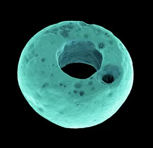
Roman glass jewellery bead, SEM
Roman glass bead. Coloured scanning electron micrograph (SEM) of a glass bead used in jewellery in Roman times

Background imageScanning Electron Microscope Collection: Ivy pollen (Hedera helix), SEM

Ivy pollen (Hedera helix), SEM
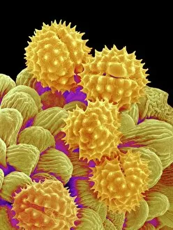
Ivy pollen (Hedera helix), coloured scanning electron micrograph (SEM). Magnification x1390 at 10cm wide

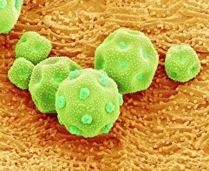
Background imageScanning Electron Microscope Collection: Philadelphia fleabane pollen grains, SEM

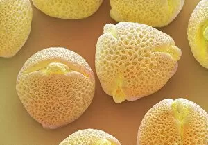
Philadelphia fleabane pollen grains, SEM
Philadelphia fleabane (Erigeron philadelphicus) pollen grains, coloured scanning electron micrograph (SEM). Pollen grains are reproductive structures produced by the male parts of flowering plants

Background imageScanning Electron Microscope Collection: Maiden hair tree stem, SEM

Maiden hair tree stem, SEM
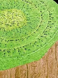
Maiden hair tree stem. Coloured scanning electron micrograph (SEM) of a section through a maidenhair tree (Ginkgo biloba) stem. At centre is the pith, which consists of soft, spongy parenchyma cells

Background imageScanning Electron Microscope Collection: Aubergine flower petal, SEM

Aubergine flower petal, SEM
Aubergine flower petal. Coloured scanning electron micrograph (SEM) of the surface of an aubergine (Solanum melongena) petal

Background imageScanning Electron Microscope Collection: Cyanobacteria, SEM

Cyanobacteria, SEM
Cyanobacteria. Coloured scanning electron micrograph (SEM) of cyanobacteria (formally known as blue-green algae, blue). Several spores (pink) are also seen

Background imageScanning Electron Microscope Collection: Eye muscle, SEM

Eye muscle, SEM
Eye muscle, coloured scanning electron micrograph (SEM). This is one of the rectus muscles of the eye. These muscles control the movement of the eyeball within the orbit (the eye socket)

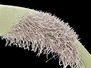
Background imageScanning Electron Microscope Collection: Banksia stem, SEM

Banksia stem, SEM
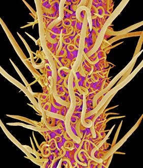
Banksia stem, coloured scanning electron micrograph (SEM). The stem is covered in fine hairs

Background imageScanning Electron Microscope Collection: Gorse stigma with pollen grains, SEM

Gorse stigma with pollen grains, SEM
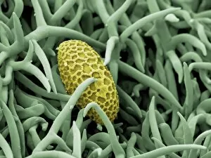
Gorse (Ulex europaeus) stigma (green) with pollen grains (yellow), coloured scanning electron micrograph (SEM). Pollen grains are reproductive structures produced by the male parts of flowering

Background imageScanning Electron Microscope Collection: Lily pollen grain on rosemary leaf, SEM

Lily pollen grain on rosemary leaf, SEM
Lily pollen grain on rosemary leaf. Coloured scanning electron micrograph (SEM) of a pollen grain (yellow) from a lily flower (Lilium sp.)

Background imageScanning Electron Microscope Collection: Gorse flower bud, SEM

Gorse flower bud, SEM
Gorse (Ulex europaeus) flower bud, coloured scanning electron micrograph (SEM). One of the petals (yellow) is seen enclosed in the flower casing (calyx)

Background imageScanning Electron Microscope Collection: Forsythia pollen grains, SEM

Forsythia pollen grains, SEM
Forsythia pollen grains arranged in a spiral, coloured scanning electron micrograph (SEM). Pollen grains are reproductive structures produced by the male parts of flowering plants

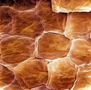
Background imageScanning Electron Microscope Collection: Skin surface, SEM

Skin surface, SEM
Skin surface. Coloured scanning electron micrograph (SEM) of squamous cells on the surface of the skin. These are flat, keratinised

Background imageScanning Electron Microscope Collection: Snake head, SEM

Snake head, SEM
Snake (suborder Serpentes) head, coloured scanning electron micrograph (SEM). The snakes forked tongue (right) is used to detect prey by picking up scent particles from the air

Background imageScanning Electron Microscope Collection: Chickweed pollen grains, SEM

Chickweed pollen grains, SEM
Chickweed pollen grains. Coloured scanning electron micrograph (SEM) of pollen grains (green) from a chickweed flower (Stellaria media)

Background imageScanning Electron Microscope Collection: Flame lily pollen, SEM

Flame lily pollen, SEM
Flame lily (Gloriosa sp.) pollen grains, coloured scanning electron micrograph (SEM). The grains have a highly sculptured outer wall (exine)

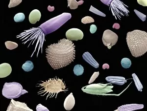
Background imageScanning Electron Microscope Collection: Mixture of flower and grass seeds, SEM

Mixture of flower and grass seeds, SEM
Mixture of flower and grass seeds, coloured scanning electron micrograph (SEM). This is a mixture of seeds for wild meadow plants. It includes flower and grass seeds

Background imageScanning Electron Microscope Collection: Calcium phosphate crystal, SEM

Calcium phosphate crystal, SEM
Calcium phosphate crystal, coloured scanning electron micrograph (SEM). Crystalline materials have their atoms placed in regular lattices that can form geometric shapes like this

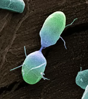
Background imageScanning Electron Microscope Collection: Salmonella bacterium dividing, SEM

Salmonella bacterium dividing, SEM
Salmonella bacterium dividing. Coloured scanning electron micrograph (SEM) of of a Salmonella bacterium dividing. The two new daughter cells are seen at upper right and lower left

Background imageScanning Electron Microscope Collection: Bacteria infecting a macrophage, SEM

Bacteria infecting a macrophage, SEM
Bacteria infecting a macrophage. Coloured scanning electron micrograph (SEM) of Mycobacterium tuberculosis bacteria (purple) infecting a macrophage white blood cell

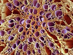
Background imageScanning Electron Microscope Collection: Xylem tissue, SEM

Xylem tissue, SEM
Xylem tissue. Coloured scanning electron micrograph (SEM) of a section through xylem tissue from a dicotyledon rootlet. Xylem vessels (purple)

Background imageScanning Electron Microscope Collection: Root of a germinating red chard seed, SEM

Root of a germinating red chard seed, SEM
Root of a germinating red chard seed. Coloured scanning electron micrograph (SEM) of a red chard seed (Beta vulgaris var. cicla) root

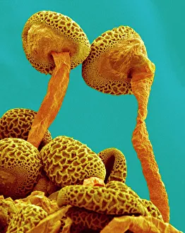
Background imageScanning Electron Microscope Collection: Pollen tubes of lily pollen, SEM

Pollen tubes of lily pollen, SEM
Pollen tubes of lily pollen, coloured scanning electron micrograph (SEM). Two pollen grains (brown) have sprouted pollen tubes (orange)

Background imageScanning Electron Microscope Collection: Germinating lily pollen, SEM

Germinating lily pollen, SEM
Germinating lily pollen. Coloured scanning electron micrograph (SEM) of lily (Lilium sp.) pollen grains that have sprouted pollen tubes. Pollen contains the male sex cells of a plant

Background imageScanning Electron Microscope Collection: Flower, SEM

Flower, SEM
Flower. Coloured scanning electron micrograph (SEM) of a flower

Background imageScanning Electron Microscope Collection: Spider lily flower stamen, SEM

Spider lily flower stamen, SEM
Spider lily flower stamen. Coloured scanning electron micrograph (SEM) of a cilia (hair-like structures) from a spider lily flower (Hymenocallis sp.) stamen

Background imageScanning Electron Microscope Collection: Leaf oil glands, SEM

Leaf oil glands, SEM
Leaf oil glands. Coloured scanning electron micrograph (SEM) of a thyme plants leaf surface (Thymus vulgaris). Two types of leaf trichomes (hairs) are seen

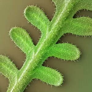
Background imageScanning Electron Microscope Collection: French lavender leaf, SEM

French lavender leaf, SEM
French lavender (Lavandula dentata) leaf. Coloured scanning electron micrograph (SEM) of a section of a French lavender leaf, showing the tooth-like structures (trichomes)

Background imageScanning Electron Microscope Collection: Grape tendril, SEM

Grape tendril, SEM
Grape tendril. Coloured scanning electron micrograph (SEM) of a curled grape (Vitis sp.) tendril. Tendrils are specialised thread-like stems or leaves that are used by climbing plants for support

Background imageScanning Electron Microscope Collection: Daisy bud, SEM

Daisy bud, SEM
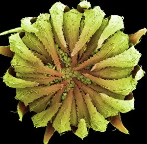
Daisy. Coloured scanning electron micrograph of a daisy bud (family Asteraceae, formerly Compositae). The protective bracts (green) cover clusters of tiny flowers, or florets

Background imageScanning Electron Microscope Collection: Zinnia flower petal, SEM

Zinnia flower petal, SEM
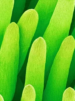
Zinnia flower petal. Coloured scanning electron micrograph (SEM) of a Zinnia sp. petal. The projections on the petal help to prevent water loss

Background imageScanning Electron Microscope Collection: Pollination, SEM

Pollination, SEM
Pollination. Coloured scanning electron micrograph (SEM) of part of a flowers pistil with pollen grains attached. The pistil is the female reproductive part of a flower

Background imageScanning Electron Microscope Collection: French lavender leaf pore, SEM

French lavender leaf pore, SEM
French lavender leaf pore. Coloured scanning electron micrograph (SEM) of an open stoma (centre, black). Stomata are pores that open and close in order to regulate gas exchange in a plant

Background imageScanning Electron Microscope Collection: Diatom alga, SEM

Diatom alga, SEM
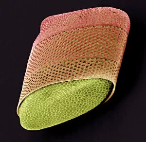
Diatom. Coloured scanning electron micrograph (SEM) of a Biddulphia sp. diatom. This is a marine planktonic unicellular alga. It has a mineralised cell wall (frustule) divided into two halves

Background imageScanning Electron Microscope Collection: Scanning Electron Micrograph (SEM): Mosquito, Female; Magnification x 55 (A4 size: 29. 7 cm width)

Scanning Electron Micrograph (SEM): Mosquito, Female; Magnification x 55 (A4 size: 29. 7 cm width)
LRDS-83 Mosquito, Female Scanning Electron Micrograph (SEM) Anopheles sp. Magnification x 55 (A4 size: 29.7 cm width) Coloured by hand to enhance natural features

Background imageScanning Electron Microscope Collection: Human chromosomes, SEM C013 / 5002

Human chromosomes, SEM C013 / 5002
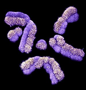
Human chromosomes. Coloured scanning electron micrograph (SEM) of human chromosomes prepared with the harlequin staining technique


All Professionally Made to Order for Quick Shipping

"Unveiling the Hidden World: Exploring with a Scanning Electron Microscope" The scanning electron microscope (SEM) is an incredible tool that allows us to delve into the microscopic realm, revealing intricate details and unlocking secrets of the unseen. With its high magnification capabilities, it offers a glimpse into fascinating aspects of life and nature. In one captivating image, we witness the beauty of gender determination as X and Y chromosomes are captured under the SEM's lens. The intricate patterns and structures within these chromosomes showcase the complexity of our genetic makeup. Moving on to another astonishing discovery, a Scanning Electron Micrograph presents us with an up-close view of a Praying Mantis at 30 times magnification. Every tiny detail becomes visible, from its delicate limbs to its mesmerizing eyes – reminding us of nature's remarkable diversity. Shifting focus to plant life, lavender pollen grains come alive in another SEM image. These minuscule particles take center stage as their unique shapes and textures become apparent under intense scrutiny. Zooming in even further reveals a Tardigrade or 'Water Bear' at an astounding 1250 times magnification. This resilient creature captures our imagination as we explore its alien-like features through this powerful imaging technique. Delving deeper into biological wonders, T lymphocytes battling cancer cells are showcased in yet another breathtaking SEM capture. This visual representation highlights the ongoing fight within our bodies against diseases like cancer – emphasizing both hope and resilience. Calcareous phytoplankton takes center stage next as we uncover their intricate forms through SEM imagery. These microscopic organisms play crucial roles in marine ecosystems while leaving us awe-inspired by their stunningly detailed structures. Transitioning to neural marvels, nerve cells reveal their intricacy under the watchful eye of an SEM. Their branching extensions create complex networks that enable communication throughout our bodies – showcasing nature's ingenuity at work. Venturing into insect realms once again, a fruit fly is immortalized under the SEM's lens.