Fraud Blocker Skip to main content

Scanning Electron Microscope Collection (page 3)

"Unveiling the Hidden World

Background imageScanning Electron Microscope Collection: Cauliflower head, SEM

Cauliflower head, SEM
Cauliflower. Coloured scanning electron micrograph (SEM) of the head of a cauliflower (Brassica oleracea botrytis). The arrangement of florets around the head is according to a Fibonacci series

Background imageScanning Electron Microscope Collection: Kidney glomerulus, SEM

Kidney glomerulus, SEM
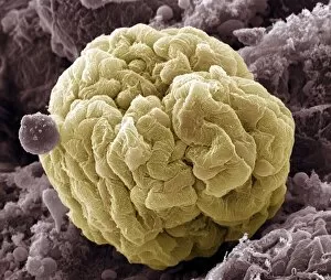
Kidney glomerulus, coloured scanning electron micrograph (SEM). Podocyte cells, which make up the surface of the glomerulus, are yellow. Magnification: x650 when printed at 10 centimetres wide

Background imageScanning Electron Microscope Collection: Carpet beetle larva, SEM

Carpet beetle larva, SEM
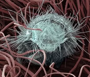
Carpet beetle larva. Coloured scanning electron micrograph (SEM) of a carpet beetle (Anthrenus sp.) larva amongst the fibres of a wool carpet

Background imageScanning Electron Microscope Collection: Macrophage cells, SEM

Macrophage cells, SEM
Macrophage cells. Coloured scanning electron micrograph (SEM) of macrophage white blood cells. Macrophages are cells of the bodys immune system

Background imageScanning Electron Microscope Collection: Ovarian follicle, SEM

Ovarian follicle, SEM
Ovarian follicle. Coloured scanning electron micrograph (SEM) of a fracture through a secondary follicle in the ovary. The oocyte (developing egg) is orange and its central nucleus is darker orange

Background imageScanning Electron Microscope Collection: Dinoflagellate plankton, SEM

Dinoflagellate plankton, SEM
Dinoflagellate plankton. Coloured scanning electron micrograph (SEM) of the calcareous (calcium-based) test (shell) of a dinoflagellate Calciodinellum sp

Background imageScanning Electron Microscope Collection: Spiny spider, SEM

Spiny spider, SEM
Spiny spider. Coloured scanning electron micrograph (SEM) of a spiny spider (Gasteracantha sp.), showing its characteristic spines (red) and large mouthparts (grey, centre left)

Background imageScanning Electron Microscope Collection: Housefly foot, SEM

Housefly foot, SEM
Housefly foot. Coloured scanning electron micrograph (SEM) of the foot of a housefly (Musca domestica). The two claws are used to grip rough surfaces

Background imageScanning Electron Microscope Collection: Trachea lining, SEM

Trachea lining, SEM
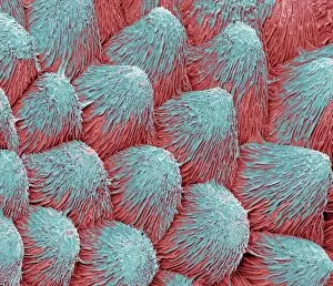
Trachea lining, coloured scanning electron micrograph (SEM). The tracheal lining contains mucus-secreting goblet cells and epithelial cells bearing hair-like cilia (blue)

Background imageScanning Electron Microscope Collection: Dandelion pollen grain, SEM

Dandelion pollen grain, SEM
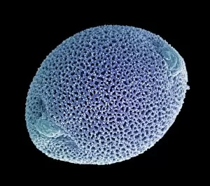
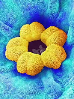
Dandelion (Taraxacum officinale) pollen grain, coloured scanning electron micrograph (SEM). Pollen grains are reproductive structures produced by the male parts of flowering plants

Background imageScanning Electron Microscope Collection: Golgi apparatus, SEM

Golgi apparatus, SEM
Golgi apparatus, coloured scanning electron micrograph (SEM). Section through a liver cell showing its Golgi apparatus (grey), a membrane-bound organelle that modifies and packages proteins

Background imageScanning Electron Microscope Collection: Goosegrass fruits, SEM

Goosegrass fruits, SEM
Goosegrass fruits. Coloured scanning electron micrograph (SEM) of the hooked fruits of a goosegrass plant (Galium aparine)

Background imageScanning Electron Microscope Collection: Synapse nerve junctions, SEM

Synapse nerve junctions, SEM
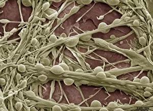
Synapse nerve junctions. Coloured scanning electron micrograph (SEM) of nerve cells showing the synapses (junctions, bulges) between them

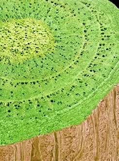
Background imageScanning Electron Microscope Collection: Silver birch twig, SEM

Silver birch twig, SEM
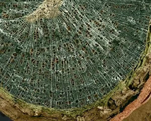
Silver birch twig. Coloured scanning electron micrograph (SEM) of a section through a silver birch (Betula pendula) twig. At the centre of the twig (top centre left) is the pith

Background imageScanning Electron Microscope Collection: Forget-me-not flower, SEM

Forget-me-not flower, SEM
Forget-me-not flower. Coloured scanning electron micrograph (SEM) of a forget-me-not (Myosotis arvensis) flower. The petals (blue) are covered by numerous projections called papillae

Background imageScanning Electron Microscope Collection: Pollen on a bee leg, SEM

Pollen on a bee leg, SEM
Pollen on a bee leg. Coloured scanning electron micrograph (SEM) of pollen grains (pale purple) on the leg of a bee (superfamily Apoidea). Pollen grains are the male sex cells of a flowering plant

Background imageScanning Electron Microscope Collection: Pseudomonas aeruginosa bacteria, SEM

Pseudomonas aeruginosa bacteria, SEM
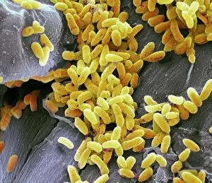
Pseudomonas aeruginosa bacteria, coloured scanning electron micrograph (SEM). These Gram-negative rod-shaped bacteria are found in soil and water, and as normal flora in the human intestine

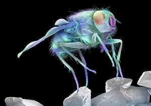
Background imageScanning Electron Microscope Collection: Fly on sugar, SEM

Fly on sugar, SEM
Fly on sugar, coloured scanning electron micrograph (SEM). Flies (order Diptera) are insects that can spread disease when they feed on human food

Background imageScanning Electron Microscope Collection: SEM of a hooks and loops fastener

SEM of a hooks and loops fastener
Coloured scanning electron micrograph (SEM)of a hooks and loops fastener. It is a two-sheet material that is used as a reversible fastener on clothing and fabrics

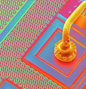
Background imageScanning Electron Microscope Collection: Microchip connecting wire, SEM

Microchip connecting wire, SEM
Micro-wire. Coloured scanning electron micrograph (SEM) of a micro-wire (yellow, often made of gold) connecting to the surface of a microchip

Background imageScanning Electron Microscope Collection: Suckling clover (Trifolium dubium), SEM C015 / 9231

Suckling clover (Trifolium dubium), SEM C015 / 9231
Suckling clover (Trifolium dubium) flower, coloured scanning electron micrograph (SEM)

Background imageScanning Electron Microscope Collection: Foraminiferan test (shell) SEM

Foraminiferan test (shell) SEM
Foraminiferan test or shell (Elphidium crispum) lateral view, coloured scanning electron micrograph (SEM). Foraminifera are single-celled protozoa which construct

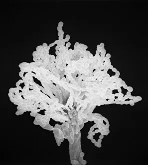
Background imageScanning Electron Microscope Collection: Aspergillus

Aspergillus
An SEM image of aspergillus in spore production (x 815 on a standard 9 cm wide print). The moulds are common in the northern hemisphere and some cause disease in humans and animals

Background imageScanning Electron Microscope Collection: Caterpillar egg

Caterpillar egg
Scanning electron microscope image of a caterpillar egg (x 90), the caterpillar emerges by chewing through the shell (x 350)

Background imageScanning Electron Microscope Collection: Blackfly antenna

Blackfly antenna
Scanning electron microscope image of a blackfly antenna (x 350). These long sensory organs feel and taste objects as well as sensing vibrations and smells (x 1.1K)

Background imageScanning Electron Microscope Collection: Anopheles gambiae, mosquito

Anopheles gambiae, mosquito
Scanning electron microscope image showing a close-up of the compound eye of a female mosquito (x 2200 on a standard 9 cm wide print)

Background imageScanning Electron Microscope Collection: Dinoflagellates, SEM

Dinoflagellates, SEM
Dinoflagellates. Coloured scanning electron micrograph (SEM) of Prorocentrum dinoflagellates (round) and Ceratium dinoflagellates (branched). Dinoflagellates are unicellular protozoans

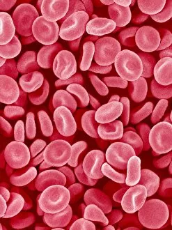
Background imageScanning Electron Microscope Collection: Red blood cells, SEM

Red blood cells, SEM
Red blood cells. Coloured scanning electron micrograph (SEM) of red blood cells (erythrocytes). Red blood cells are biconcave, disc-shaped cells that transport oxygen from the lungs to body cells

Background imageScanning Electron Microscope Collection: Diatom frustule, SEM

Diatom frustule, SEM
Diatom frustule. Coloured scanning electron micrograph (SEM) of the frustule of a Pinnularia sp. diatom. The diatoms are a group of photosynthetic, single-celled algae containing about 10

Background imageScanning Electron Microscope Collection: Buttercup flower, SEM

Buttercup flower, SEM
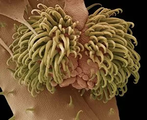
Buttercup flower (Ranunculus sp.), coloured scanning electron micrograph (SEM). The pistil (centre) is surrounded by the stamens (yellow)

Background imageScanning Electron Microscope Collection: Diatom cell wall, SEM

Diatom cell wall, SEM
Diatom cell wall. Coloured scanning electron micrograph (SEM) of the pattern of small holes, called striae, in the cell wall of a diatom alga. This is a Coscinodiscus sp. diatom

Background imageScanning Electron Microscope Collection: Gecko foot, SEM

Gecko foot, SEM
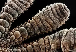
Gecko foot. Coloured scanning electron micrograph (SEM) of the underside of a geckos foot (Tarentola mauritanica). The foot is covered with ridges and microscopic hairs

Background imageScanning Electron Microscope Collection: Fruit fly, SEM Z340 / 0700

Fruit fly, SEM Z340 / 0700
Fruit fly. Coloured scanning electron micrograph (SEM) of the head of a fruit fly (Drosophila busckii). Its two compound eyes (red) are seen on either side of the head

Background imageScanning Electron Microscope Collection: Oxytricha ciliate protozoan, SEM C019 / 0253

Oxytricha ciliate protozoan, SEM C019 / 0253
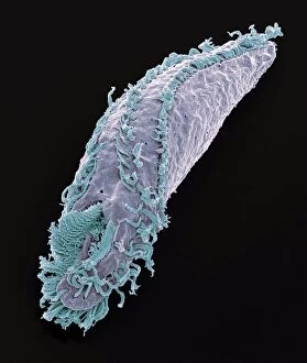
Oxytricha sp. ciliate protozoan, coloured scanning electron micrograph (SEM). Oxytricha is a tiny single-celled aquatic organism

Background imageScanning Electron Microscope Collection: Butterfly wing scales

Butterfly wing scales. Coloured scanning electron micrograph (SEM) of scales from the topside of a wing of an aega morpho (Morpho aega) butterfly

Background imageScanning Electron Microscope Collection: T2 cell culture

T2 cell culture
Scanning electron microscope image showing a T2 cell culture (x 4K)

Background imageScanning Electron Microscope Collection: Mouse malaria parasite, SEM

Mouse malaria parasite, SEM
Mouse malaria parasite. Coloured scanning electron micrograph (SEM) of a Plasmodium berghei protozoan (yellow) and red blood cells. P. berghei is the parasite that causes malaria in mice

Background imageScanning Electron Microscope Collection: Orchid petal, SEM

Orchid petal, SEM
Orchid (Phalaenopsis sp.) petal, coloured scanning electron micrograph (SEM). Magnification: x450 when printed at 10 centimetres wide

Background imageScanning Electron Microscope Collection: Stem cell, SEM

Stem cell, SEM
Stem cell, coloured scanning electron micrograph (SEM). Stem cells can differentiate into any other cell type. There are three main types of mammalian stem cell: embryonic stem cells

Background imageScanning Electron Microscope Collection: Stem cells, SEM

Stem cells, SEM
Stem cells, coloured scanning electron micrograph (SEM). Stem cells can differentiate into any other cell type. There are three main types of mammalian stem cell: embryonic stem cells

Background imageScanning Electron Microscope Collection: Mycobacterium chelonae bacteria, SEM

Mycobacterium chelonae bacteria, SEM
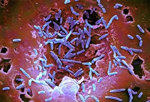
Mycobacterium chelonae bacteria, coloured scanning electron micrograph (SEM). These Gram-positive rod-shaped bacteria are found in soil and water

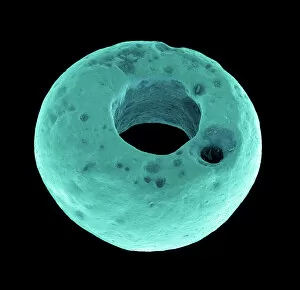
Background imageScanning Electron Microscope Collection: Roman glass jewellery bead, SEM

Roman glass jewellery bead, SEM
Roman glass bead. Coloured scanning electron micrograph (SEM) of a glass bead used in jewellery in Roman times

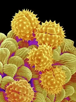
Background imageScanning Electron Microscope Collection: Ivy pollen (Hedera helix), SEM

Ivy pollen (Hedera helix), SEM
Ivy pollen (Hedera helix), coloured scanning electron micrograph (SEM). Magnification x1390 at 10cm wide

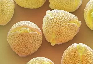
Background imageScanning Electron Microscope Collection: Philadelphia fleabane pollen grains, SEM

Philadelphia fleabane pollen grains, SEM
Philadelphia fleabane (Erigeron philadelphicus) pollen grains, coloured scanning electron micrograph (SEM). Pollen grains are reproductive structures produced by the male parts of flowering plants

Background imageScanning Electron Microscope Collection: Maiden hair tree stem, SEM

Maiden hair tree stem, SEM
Maiden hair tree stem. Coloured scanning electron micrograph (SEM) of a section through a maidenhair tree (Ginkgo biloba) stem. At centre is the pith, which consists of soft, spongy parenchyma cells

Background imageScanning Electron Microscope Collection: Aubergine flower petal, SEM

Aubergine flower petal, SEM
Aubergine flower petal. Coloured scanning electron micrograph (SEM) of the surface of an aubergine (Solanum melongena) petal

Background imageScanning Electron Microscope Collection: Cyanobacteria, SEM

Cyanobacteria, SEM
Cyanobacteria. Coloured scanning electron micrograph (SEM) of cyanobacteria (formally known as blue-green algae, blue). Several spores (pink) are also seen

Background imageScanning Electron Microscope Collection: Eye muscle, SEM

Eye muscle, SEM
Eye muscle, coloured scanning electron micrograph (SEM). This is one of the rectus muscles of the eye. These muscles control the movement of the eyeball within the orbit (the eye socket)


All Professionally Made to Order for Quick Shipping

"Unveiling the Hidden World: Exploring with a Scanning Electron Microscope" The scanning electron microscope (SEM) is an incredible tool that allows us to delve into the microscopic realm, revealing intricate details and unlocking secrets of the unseen. With its high magnification capabilities, it offers a glimpse into fascinating aspects of life and nature. In one captivating image, we witness the beauty of gender determination as X and Y chromosomes are captured under the SEM's lens. The intricate patterns and structures within these chromosomes showcase the complexity of our genetic makeup. Moving on to another astonishing discovery, a Scanning Electron Micrograph presents us with an up-close view of a Praying Mantis at 30 times magnification. Every tiny detail becomes visible, from its delicate limbs to its mesmerizing eyes – reminding us of nature's remarkable diversity. Shifting focus to plant life, lavender pollen grains come alive in another SEM image. These minuscule particles take center stage as their unique shapes and textures become apparent under intense scrutiny. Zooming in even further reveals a Tardigrade or 'Water Bear' at an astounding 1250 times magnification. This resilient creature captures our imagination as we explore its alien-like features through this powerful imaging technique. Delving deeper into biological wonders, T lymphocytes battling cancer cells are showcased in yet another breathtaking SEM capture. This visual representation highlights the ongoing fight within our bodies against diseases like cancer – emphasizing both hope and resilience. Calcareous phytoplankton takes center stage next as we uncover their intricate forms through SEM imagery. These microscopic organisms play crucial roles in marine ecosystems while leaving us awe-inspired by their stunningly detailed structures. Transitioning to neural marvels, nerve cells reveal their intricacy under the watchful eye of an SEM. Their branching extensions create complex networks that enable communication throughout our bodies – showcasing nature's ingenuity at work. Venturing into insect realms once again, a fruit fly is immortalized under the SEM's lens.