Fraud Blocker Skip to main content

Scanning Electron Microscope Collection (page 7)

"Unveiling the Hidden World

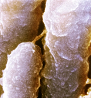
Background imageScanning Electron Microscope Collection: Inner ear hairs, SEM

Inner ear hairs, SEM
Inner ear hairs. Coloured scanning electron micrograph (SEM) of stereocilia, sensory hair-like projections, from hair cells of the cochlea of the inner ear

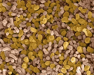
Background imageScanning Electron Microscope Collection: Yeast cells, SEM

Yeast cells, SEM
Yeast cells. Coloured scanning electron micrograph (SEM) of cells of bakers yeast (Saccharomyces cerevisiae) from part of a dried, commercial yeast pellet

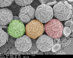
Background imageScanning Electron Microscope Collection: Emiliania huxleyi coccolithophores

Emiliania huxleyi coccolithophores collected from a bloom in the SW Approaches to the English Channel in June 2004. Date: 2004

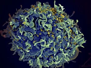
Background imageScanning Electron Microscope Collection: Scanning electron micrograph of HIV particles infecting a human H9 T cell

Scanning electron micrograph of HIV particles infecting a human H9 T cell

Background imageScanning Electron Microscope Collection: Coxiella burnetii, the bacteria that causes Q Fever

Coxiella burnetii, the bacteria that causes Q Fever. A dry fracture of a Vero cell exposing the contents of a vacuole where Coxiella burnetii are busy growing

Background imageScanning Electron Microscope Collection: HIV infected macrophage, SEM C018 / 8598

HIV infected macrophage, SEM C018 / 8598
HIV infected macrophage. Coloured ion-abrasion scanning electron micrograph (IA-SEM) of a macrophage white blood cell infected with human immunodeficiency virus (HIV, red)

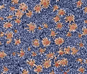
Background imageScanning Electron Microscope Collection: Nasal lining, SEM

Nasal lining, SEM
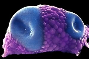
Nasal lining. Coloured scanning electron micrograph (SEM) of the olfactory epithelium that lines the nasal cavity, showing olfactory cells (red) surrounded by numerous cilia (hair-like projections)

Background imageScanning Electron Microscope Collection: Intestinal villi, SEM

Intestinal villi, SEM
Intestinal villi. Coloured scanning electron micrograph (SEM) of a section through a part of the small intestine, showing the villi that cover its inner surface

Background imageScanning Electron Microscope Collection: Butterfly eye, SEM C016 / 9382

Butterfly eye, SEM C016 / 9382
Butterfly eye. Coloured scanning electron micrograph (SEM) of the compound eye (blue) of a butterfly, showing the individual lenses (ommatidia)

Background imageScanning Electron Microscope Collection: Hydra budding, SEM

Hydra budding, SEM
Hydra budding. Coloured scanning electron micrograph (SEM) of a Hydra sp. freshwater hydrozoan budding. Hydra commonly reproduce by budding - a form of asexual reproduction in which a new organism

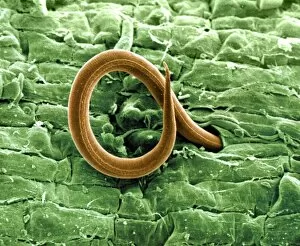
Background imageScanning Electron Microscope Collection: Root-knot nematode larva, SEM

Root-knot nematode larva, SEM
Root-knot nematode larva. Coloured scanning electron micrograph (SEM) of the whiplike larva of the root-knot nematode Meloidogyne incognita. Here, it is penetrating a tomato root

Background imageScanning Electron Microscope Collection: Trachea lining, SEM C015 / 9936

Trachea lining, SEM C015 / 9936
Trachea lining. Coloured scanning electron micrograph (SEM) of a section through the lining of the trachea (wind pipe). The trachea links the larynx (voice box) to the lungs

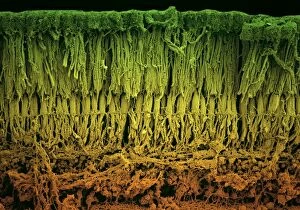
Background imageScanning Electron Microscope Collection: Retina layers, SEM C018 / 0520

Retina layers, SEM C018 / 0520
Retina layers, coloured scanning electron micrograph (SEM). The retina of the human eye is found on the inside of the eyeball and is formed from a number of layers

Background imageScanning Electron Microscope Collection: Ant anus, SEM C018 / 0550

Ant anus, SEM C018 / 0550
Ant anus, coloured scanning electron micrograph (SEM). Ants excrete bodily waste through a single orifice, their anus, which is located at the tip of the abdomen

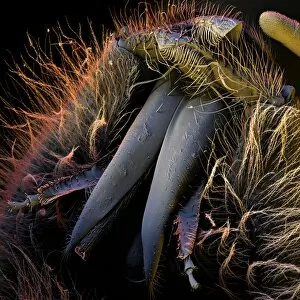
Background imageScanning Electron Microscope Collection: Bee head, SEM C018 / 0546

Bee head, SEM C018 / 0546
Bee head, coloured scanning electron micrograph (SEM). This is the head of a small bumblebee. Flying insects often have two sets of eyes, coloured pink here

Background imageScanning Electron Microscope Collection: Rod and cone cells of the eye, SEM C014 / 4864

Rod and cone cells of the eye, SEM C014 / 4864
Rod and cone cells of the eye. Coloured scanning electron micrograph (SEM) of rod and cone cells in the retina of a mammalian eye

Background imageScanning Electron Microscope Collection: Hydra budding C014 / 4856

Hydra budding C014 / 4856
Hydra budding. Coloured scanning electron micrograph (SEM) of a Hydra viridis freshwater hydrozoan. Hydra viridis commonly reproduces by budding, as seen here

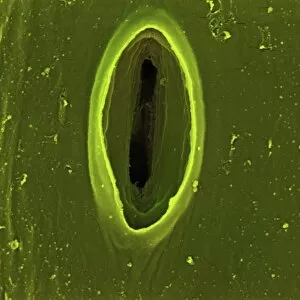
Background imageScanning Electron Microscope Collection: Chives leaf stoma, SEM C016 / 8060

Chives leaf stoma, SEM C016 / 8060
Chives leaf stoma. Coloured scanning electron micrograph (SEM) of a stoma (oval, centre) on the surface of a leaf from a chives (Allium schoenoprasum) plant

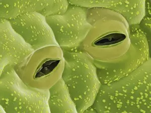
Background imageScanning Electron Microscope Collection: Coltsfoot leaf stomata, SEM C016 / 8052

Coltsfoot leaf stomata, SEM C016 / 8052
Coltsfoot leaf stomata. Coloured scanning electron micrograph (SEM) of two stomata (round) on the surface of a leaf from a coltsfoot (Tussilago farfara) plant

Background imageScanning Electron Microscope Collection: Honey bee mouth, SEM C016 / 8004

Honey bee mouth, SEM C016 / 8004
Honey bee mouth. Coloured scanning electron micrograph (SEM) of the mouth a honey bee (Apis sp.), showing the hairs that cover its body. Magnification: x37, when printed 10 centimetres high

Background imageScanning Electron Microscope Collection: Propionibacterium acnes C014 / 0631

Propionibacterium acnes C014 / 0631
Propionibacterium acnes bacteria, coloured scanning electron micrograph (SEM). This bacteria is found deep in the hair follicles and pores of the skin

Background imageScanning Electron Microscope Collection: Potato Leaf Stomata (SEM)

Potato Leaf Stomata (SEM)
Potato Stomata. Scanning electron micrographs (SEM) of open stomata on a potato leaf (Solanum tuberosum). Stomata are pores that open and close in order to regulate gas exchange in a plant

Background imageScanning Electron Microscope Collection: Skin cancer cell, SEM C016 / 4764

Skin cancer cell, SEM C016 / 4764
Skin cancer cell. Coloured ion-abrasion scanning electron micrograph (IA-SEM) of a melanoma cell. The large dark sphere is the cell nucleus, which contains the cells genetic information

Background imageScanning Electron Microscope Collection: White blood cells and platelets, SEM C016 / 3099

White blood cells and platelets, SEM C016 / 3099
White blood cells and platelets. Coloured scanning electron micrograph (SEM) of platelets (green) with a variety of different white blood cells (leucocytes, yellow)

Background imageScanning Electron Microscope Collection: White blood cells and platelets, SEM C016 / 3098

White blood cells and platelets, SEM C016 / 3098
White blood cells and platelets. Coloured scanning electron micrograph (SEM) of platelets (purple) with a variety of different white blood cells (leucocytes, blue)

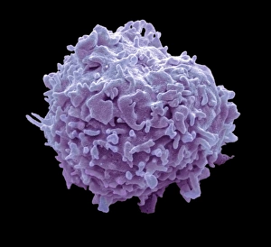
Background imageScanning Electron Microscope Collection: Monocyte white blood cell, SEM C016 / 3089

Monocyte white blood cell, SEM C016 / 3089
Monocyte white blood cell. Scanning electron micrograph (SEM) of a monocyte white blood cell. Monocytes, like all white blood cells, are part of the human bodys immune system

Background imageScanning Electron Microscope Collection: Trachea lining, SEM C013 / 7126

Trachea lining, SEM C013 / 7126
Trachea lining. Coloured scanning electron micrograph (SEM) of the lining of a trachea (wind pipe), showing the cilia (hair-like) and goblet cells (orange). The trachea links the larynx to the lungs

Background imageScanning Electron Microscope Collection: Trachea lining, SEM C013 / 7122

Trachea lining, SEM C013 / 7122
Trachea lining. Coloured scanning electron micrograph (SEM) of a section through the wall of a trachea (wind pipe). The trachea links the larynx to the lungs

Background imageScanning Electron Microscope Collection: Common salt crystals SEM

Common salt crystals SEM
Coloured scanning electron micrograph (SEM) of common salt, sodium chloride, recrystallised from distilled water. Magnification x225 at 10cm wide

Background imageScanning Electron Microscope Collection: Tardigrade or water bear egg SEM

Tardigrade or water bear egg SEM
Water bear (Macrobiotus sp.) egg, coloured scanning electron micrograph (SEM). Water bears (or tardigrades) are tiny invertebrates that live in aquatic and semi-aquatic habitats such as lichen

Background imageScanning Electron Microscope Collection: Distended fat cell, SEM C013 / 5015

Distended fat cell, SEM C013 / 5015
Distended fat cell. Coloured scanning electron micrograph (SEM) of a large adipocyte (fat cell, orange) distended by fat droplets (green)

Background imageScanning Electron Microscope Collection: Distended fat cell, SEM C013 / 5013

Distended fat cell, SEM C013 / 5013
Distended fat cell. Coloured scanning electron micrograph (SEM) of a large adipocyte (fat cell, blue) distended by fat droplets (purple)

Background imageScanning Electron Microscope Collection: Human Red Blood Cells, SEM

Human Red Blood Cells, SEM
Human red blood cells, coloured composite scanning electron micrograph (SEM)

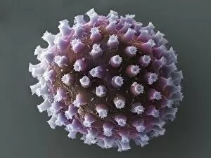
Background imageScanning Electron Microscope Collection: Lavender pollen grain, SEM

Lavender pollen grain, SEM
Lavender pollen grain. Coloured scanning electron micrograph (SEM) of a French lavender (Lavandula dentata) flower, with an enlarged section showing a pollen grain (yellow) amongst the petal (papilla)

Background imageScanning Electron Microscope Collection: Red blood cell, SEM

Red blood cell, SEM
Red blood cell, coloured scanning electron micrograph (SEM). Red blood cells (erythrocytes) are carriers of oxygen and carbon dioxide

Background imageScanning Electron Microscope Collection: Turmeric powder, SEM

Turmeric powder, SEM
Turmeric powder, coloured scanning electron micrograph (SEM). Turmeric is a deep yellow spice made from the dried and ground rhizomes of turmeric (Curcuma longa)

Background imageScanning Electron Microscope Collection: Synthetic Mycoplasma bacteria, SEM C013 / 4777

Synthetic Mycoplasma bacteria, SEM C013 / 4777
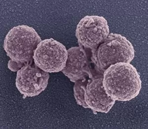
Synthetic Mycoplasma bacteria. Coloured scanning electron micrograph (SEM) of Mycoplasma mycoides JCVI-syn1.0 cells. This was the first self-replicating bacterium controlled by a synthetic genome

Background imageScanning Electron Microscope Collection: Sea salt

Sea salt
A scanning electron microscope (SEM) image of sea salt, artificially coloured by computer

Background imageScanning Electron Microscope Collection: Fragaria sp. strawberry

Fragaria sp. strawberry
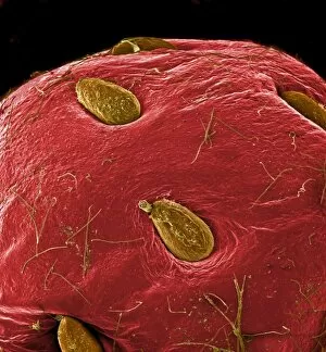
A scanning electron microscope (SEM) image of a strawberry (Fragaria sp.), artificially coloured by computer

Background imageScanning Electron Microscope Collection: Tyrophagus casei, cheese mite

Tyrophagus casei, cheese mite
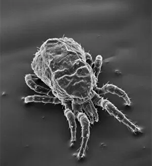
Scanning electron microscope image of a cheese mite (x 170). These creatures are generally considered to be a pest, however they are added to Altenburger cheese to give it flavour

Background imageScanning Electron Microscope Collection: SEM of echinoderm steroem

SEM of echinoderm steroem
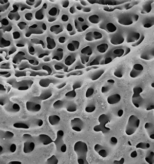
A SEM of an example of stereom of an echinoderm (phylum which consists of 5 classes including starfish). Stereom is the structure formed by the fine networks of calcium carbonate which constitute

Background imageScanning Electron Microscope Collection: Dermanyssus gallinae, red or poultry mite

Dermanyssus gallinae, red or poultry mite
Scanning electron microscope image of the red or poutry mite. Adults appear red when engorged with blood, but otherwise are black, grey or white. Females are about 1mm long

Background imageScanning Electron Microscope Collection: Ceratodon purpureus, ceratodon moss spore capsule

Ceratodon purpureus, ceratodon moss spore capsule
Scanning electron microscope (SEM) image of a ceratodon moss spore capsule (x 650 on a standard 9 cm wide print)

Background imageScanning Electron Microscope Collection: Foraminifer

Foraminifer
Scanning electron microscope (SEM) image of a foraminifer - a single celled organism

Background imageScanning Electron Microscope Collection: Dermatophagoides pteronyssius, dust mite

Dermatophagoides pteronyssius, dust mite
Scanning electron microscope image showing a dust mite (x 250 on standard 9cm wide print). This image has been artificially coloured by a computer

Background imageScanning Electron Microscope Collection: Marine Diatoms

Marine Diatoms
LRMC-3 Scanning Electron Micrograph (SEM): Marine Diatoms Magnification x2400 (when printed A4, 29.7 cm cm wide) David McCarthy and Dae Sasitorn / ardea.com Last Refuge contact details

Background imageScanning Electron Microscope Collection: Silicoflagellate

Silicoflagellate
LRDS-297 Scanning Electron Micrograph (SEM): Silicoflagellate, Skeleton bearing stage of Dictyocha speculum Magnification x3

Background imageScanning Electron Microscope Collection: Pipistrelle bat ear, SEM

Pipistrelle bat ear, SEM
Pipistrelle bat ear. Coloured scanning electron micrograph (SEM) of the ear of a pipistrelle bat (Pipistrellus pipistrellus)


All Professionally Made to Order for Quick Shipping

"Unveiling the Hidden World: Exploring with a Scanning Electron Microscope" The scanning electron microscope (SEM) is an incredible tool that allows us to delve into the microscopic realm, revealing intricate details and unlocking secrets of the unseen. With its high magnification capabilities, it offers a glimpse into fascinating aspects of life and nature. In one captivating image, we witness the beauty of gender determination as X and Y chromosomes are captured under the SEM's lens. The intricate patterns and structures within these chromosomes showcase the complexity of our genetic makeup. Moving on to another astonishing discovery, a Scanning Electron Micrograph presents us with an up-close view of a Praying Mantis at 30 times magnification. Every tiny detail becomes visible, from its delicate limbs to its mesmerizing eyes – reminding us of nature's remarkable diversity. Shifting focus to plant life, lavender pollen grains come alive in another SEM image. These minuscule particles take center stage as their unique shapes and textures become apparent under intense scrutiny. Zooming in even further reveals a Tardigrade or 'Water Bear' at an astounding 1250 times magnification. This resilient creature captures our imagination as we explore its alien-like features through this powerful imaging technique. Delving deeper into biological wonders, T lymphocytes battling cancer cells are showcased in yet another breathtaking SEM capture. This visual representation highlights the ongoing fight within our bodies against diseases like cancer – emphasizing both hope and resilience. Calcareous phytoplankton takes center stage next as we uncover their intricate forms through SEM imagery. These microscopic organisms play crucial roles in marine ecosystems while leaving us awe-inspired by their stunningly detailed structures. Transitioning to neural marvels, nerve cells reveal their intricacy under the watchful eye of an SEM. Their branching extensions create complex networks that enable communication throughout our bodies – showcasing nature's ingenuity at work. Venturing into insect realms once again, a fruit fly is immortalized under the SEM's lens.