Canvas Print > Popular Themes > Human Body
Canvas Print : Nerve and glial cells, light micrograph


![]()
Canvas Prints from Science Photo Library
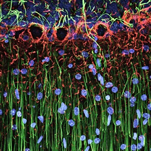
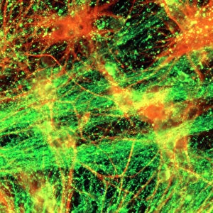
Nerve and glial cells, light micrograph
Nerve and glial cells, fluorescence light micrograph. These are neural stem cells that have differentiated into neurons (nerve cells, blue) and glial cells (support cells, red). The branching processes from the neurons are called dendrites. Fluorescent markers have been used to highlight proteins. The proteins stained here are beta III-tubulin (blue), a cytoskeleton element found in neurons, and GFAP (glial fibrillary acidic protein, red), forming the cytoskeleton of the glial cells. This sample is from rat tissue
Science Photo Library features Science and Medical images including photos and illustrations
Media ID 10948201
© DANIEL SCHROEN, CELL APPLICATIONS INC/SCIENCE PHOTO LIBRARY
Animal Body Astrocyte Astrocytes Cell Biology Cellular Cytoskeletal Cytoskeleton Fluorescence Fluorescence Micrograph Fluorescing Gfap Glial Cell Glial Fibrillary Acidic Protein Nerve Nerve Cell Neuron Neurone Neurones Neurons Nobody Proteins Stains Tubulin Brain Cells Light Micrograph Light Microscope Nervous System Neurological Neurology Protein
20"x16" (50x40cm) Canvas Print
Introducing the Media Storehouse Canvas Prints featuring the captivating image "Nerve and Glial Cells" by Daniel Schroen from Science Photo Library. This mesmerizing light micrograph showcases the intricate beauty of neural stem cells as they transform into neurons, represented in a brilliant blue hue, and glial cells, depicted in vibrant red. Our high-quality canvas prints bring this stunning scientific discovery into your home or office, adding an element of intellectual sophistication and conversation-starting intrigue. Each print is expertly crafted using premium materials to ensure long-lasting vibrancy and a rich, textured finish. Elevate your space with the Media Storehouse Canvas Prints and immerse yourself in the wonders of science.
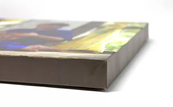
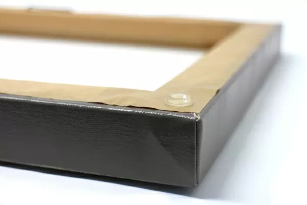
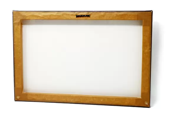
Ready to hang Premium Gloss Canvas Print. Our archival quality canvas prints are made from Polyester and Cotton mix and stretched over a 1.25" (32mm) kiln dried knot free wood stretcher bar. Packaged in a plastic bag and secured to a cardboard insert for transit.
Canvas Prints add colour, depth and texture to any space. Professionally Stretched Canvas over a hidden Wooden Box Frame and Ready to Hang
Estimated Product Size is 50.8cm x 40.6cm (20" x 16")
These are individually made so all sizes are approximate
Artwork printed orientated as per the preview above, with landscape (horizontal) orientation to match the source image.
EDITORS COMMENTS
This print showcases the intricate world of nerve and glial cells, captured through a fluorescence light micrograph. The neural stem cells in this image have undergone differentiation, transforming into neurons (nerve cells) depicted in blue, as well as glial cells (support cells) shown in red. The dendrites extending from the neurons are responsible for transmitting electrical signals throughout the nervous system. To enhance visibility and highlight specific proteins, fluorescent markers were employed during sample preparation. In this particular image, beta III-tubulin is stained blue to emphasize its presence within neuronal cytoskeletons. Meanwhile, GFAP (glial fibrillary acidic protein), which forms the structural framework of glial cells' cytoskeletons, appears vibrant red. It's important to note that this sample originates from rat tissue and offers valuable insights into cellular biology and neurology research. By studying these fundamental building blocks of our nervous system – astrocytes, neurones, differentiated stem cells – scientists gain a deeper understanding of how our brains function. The photographer behind this remarkable image is Daniel Schroen from Cell Applications Inc/Science Photo Library. This visually striking photograph not only captures the beauty found within biological structures but also serves as a testament to human curiosity and scientific exploration in unraveling the mysteries of life itself.
MADE IN THE UK
Safe Shipping with 30 Day Money Back Guarantee
FREE PERSONALISATION*
We are proud to offer a range of customisation features including Personalised Captions, Color Filters and Picture Zoom Tools
SECURE PAYMENTS
We happily accept a wide range of payment options so you can pay for the things you need in the way that is most convenient for you
* Options may vary by product and licensing agreement. Zoomed Pictures can be adjusted in the Basket.